
Overview
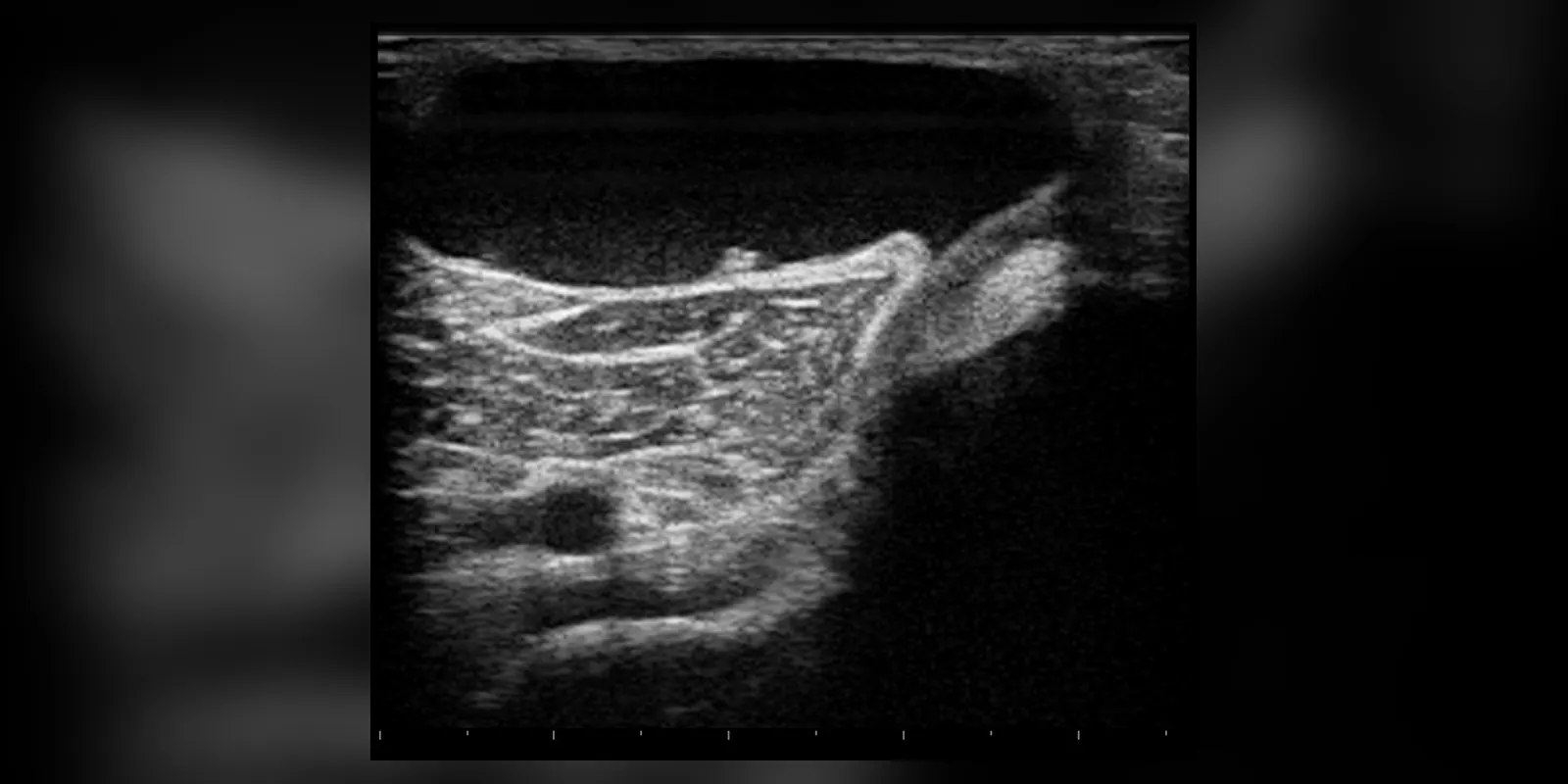
A Baker's cyst, also called a popliteal cyst, is a fluid-filled swelling at the back of the knee. It forms when joint fluid from inside the knee bulges into a small sac (bursa) behind the knee. It is named after the surgeon William Baker, who described it in the nineteenth century.
Baker's cysts are usually caused by another knee problem, such as osteoarthritis, rheumatoid arthritis or a meniscus (cartilage) tear, which makes the knee produce extra fluid. Many cause no symptoms, while others cause a feeling of tightness, swelling or pain behind the knee, especially when bending or straightening the leg. Occasionally a cyst bursts, causing sudden calf pain and swelling that can look like a blood clot. Treatment focuses on the underlying knee problem, and many cysts settle on their own.
A Baker's cyst is not a tumour and is not dangerous in itself. It is the knee's way of storing extra fluid, so it often grows and shrinks as the underlying knee problem flares and settles.
When to get urgent help
Seek urgent care for sudden calf pain and swelling, especially with redness or warmth, to rule out a blood clot, or for a hot, red, painful knee with fever.
Causes and risk factors
- Osteoarthritis of the knee, the most common cause in adults
- Rheumatoid arthritis and other inflammatory arthritis
- Meniscus tears and other cartilage damage
- Gout
- Knee injury
- In children: often no underlying cause, and the cyst usually disappears on its own
How it develops (pathophysiology)
Normal knee anatomy
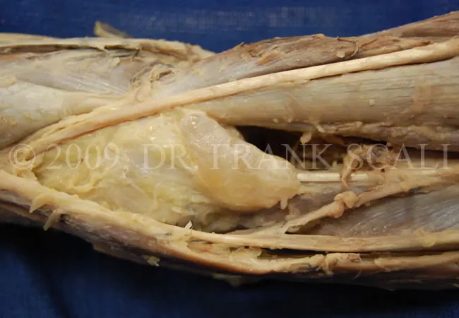
The knee joint is surrounded by a capsule lined with synovium, which produces synovial fluid to lubricate the joint. Around the knee are several bursae, small fluid-filled sacs that reduce friction between tendons, muscles and bone. At the back of the knee, the gastrocnemius-semimembranosus bursa lies between the tendons of the calf muscle (gastrocnemius) and a hamstring muscle (semimembranosus). In many adults, this bursa connects to the knee joint through a small opening.
What goes wrong
- Excess joint fluid: arthritis, cartilage tears and inflammation make the synovium produce more fluid (an effusion).
- One-way valve: the opening between the knee joint and the bursa often acts like a valve. When the knee bends, pressure inside the joint pushes fluid into the bursa; when it straightens, the opening closes, trapping fluid.
- Cyst formation: fluid accumulates in the bursa, which stretches and enlarges into a cyst behind the knee.
- Rupture: if pressure rises suddenly, the cyst can burst, releasing fluid down between the calf muscles. This causes inflammation, pain and swelling in the calf, mimicking a deep vein thrombosis.
- Compression: a large cyst may press on nearby veins or nerves, rarely causing leg swelling or numbness.
Why the symptoms happen
- Swelling behind the knee reflects fluid in the cyst.
- Tightness or stiffness, especially when fully bending or straightening, occurs because the cyst is squeezed between tendons.
- Pain may come from the cyst itself or from the underlying arthritis or cartilage damage.
- Sudden calf pain, swelling and redness occur with rupture.
How treatment acts on the mechanism
Treating the underlying knee condition reduces fluid production, so the cyst often shrinks. Steroid injections into the knee reduce inflammation and fluid. Aspiration (draining with a needle) gives temporary relief. Surgery to repair cartilage tears or treat arthritis addresses the cause, and cyst removal is rarely needed.
Types
- Primary Baker's cyst: no underlying knee problem, common in children
- Secondary Baker's cyst: linked to arthritis or a cartilage tear, common in adults
- Ruptured Baker's cyst
Signs and symptoms
- Swelling behind the knee, which may feel like a water-filled balloon
- Tightness or stiffness behind the knee
- Pain when bending or straightening the knee
- Swelling more noticeable when standing
- Symptoms of the underlying knee problem, such as knee pain, clicking or locking
- If ruptured: sudden sharp pain in the calf, swelling and redness, sometimes with bruising around the ankle
Some people notice the swelling only when they kneel, squat or stand for long periods. Many small cysts are found by chance on a scan done for another reason.
How it is diagnosed
- Examination of the knee and back of the leg
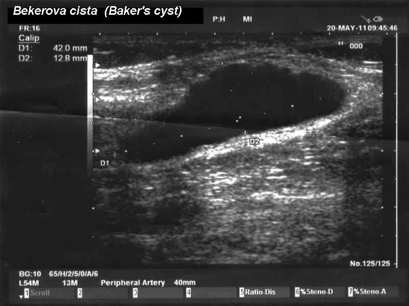
- Ultrasound, which confirms a fluid-filled cyst and can help exclude a blood clot
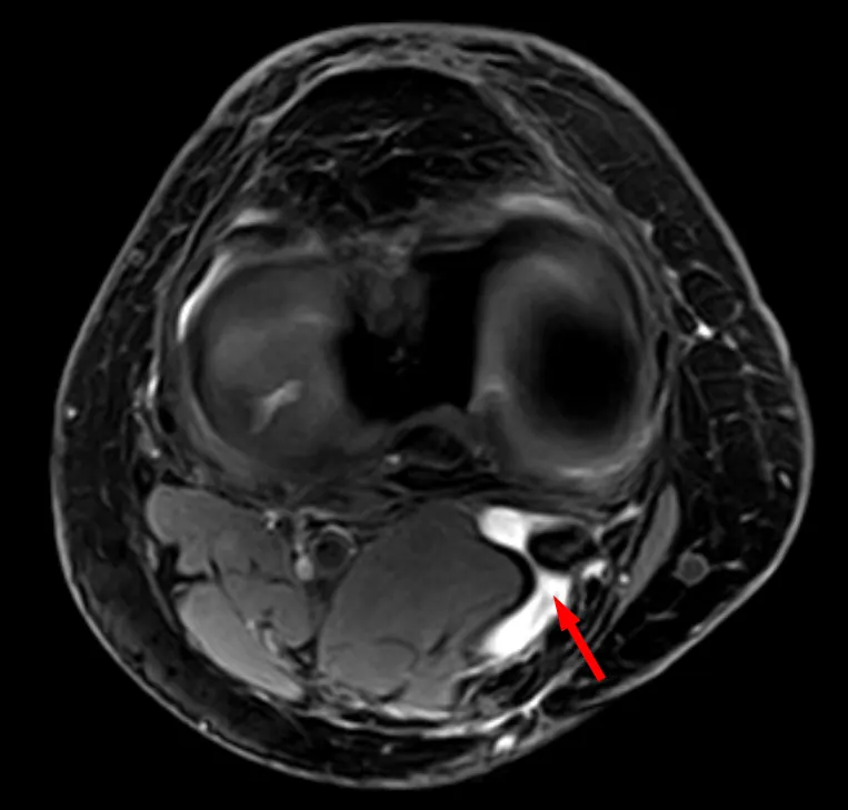
- MRI to look for cartilage tears or other causes
- X-ray to look for arthritis
- Doppler ultrasound to exclude deep vein thrombosis if calf swelling occurs
Possible complications
- Rupture, causing calf pain and swelling
- Compartment syndrome (very rare)
- Compression of veins causing leg swelling or blood clots (rare)
- Infection (rare)
Goals of treatment
Treatment aims to relieve symptoms and treat the underlying knee condition.
Medicines
- Pain relief: paracetamol or anti-inflammatories
- Steroid injection into the knee joint
- Medicines for underlying arthritis, such as treatment for rheumatoid arthritis or gout
Other treatments
- Rest and elevation
- Ice packs for swelling
- Compression bandage or support
- Physiotherapy to strengthen muscles and improve knee movement
- Aspiration of the cyst, sometimes guided by ultrasound
- Arthroscopic surgery to treat cartilage tears
- Knee replacement for severe arthritis
- Surgical removal of the cyst in rare cases
Living with it and prevention
- Manage arthritis and knee injuries early
- Keep a healthy weight to reduce knee strain
- Strengthen thigh and calf muscles
- Avoid activities that cause knee pain and swelling
Monitoring and follow-up
Cysts often settle over months. Review is needed if swelling persists, increases, or symptoms change.
Outlook
Most Baker's cysts improve when the underlying knee condition is treated. Some persist or return. In children, they usually disappear without treatment.
Special considerations
- Children: often resolve on their own; usually no surgery needed
- Older adults with arthritis: cysts are common
- People with calf swelling: blood clots must be excluded