
Overview
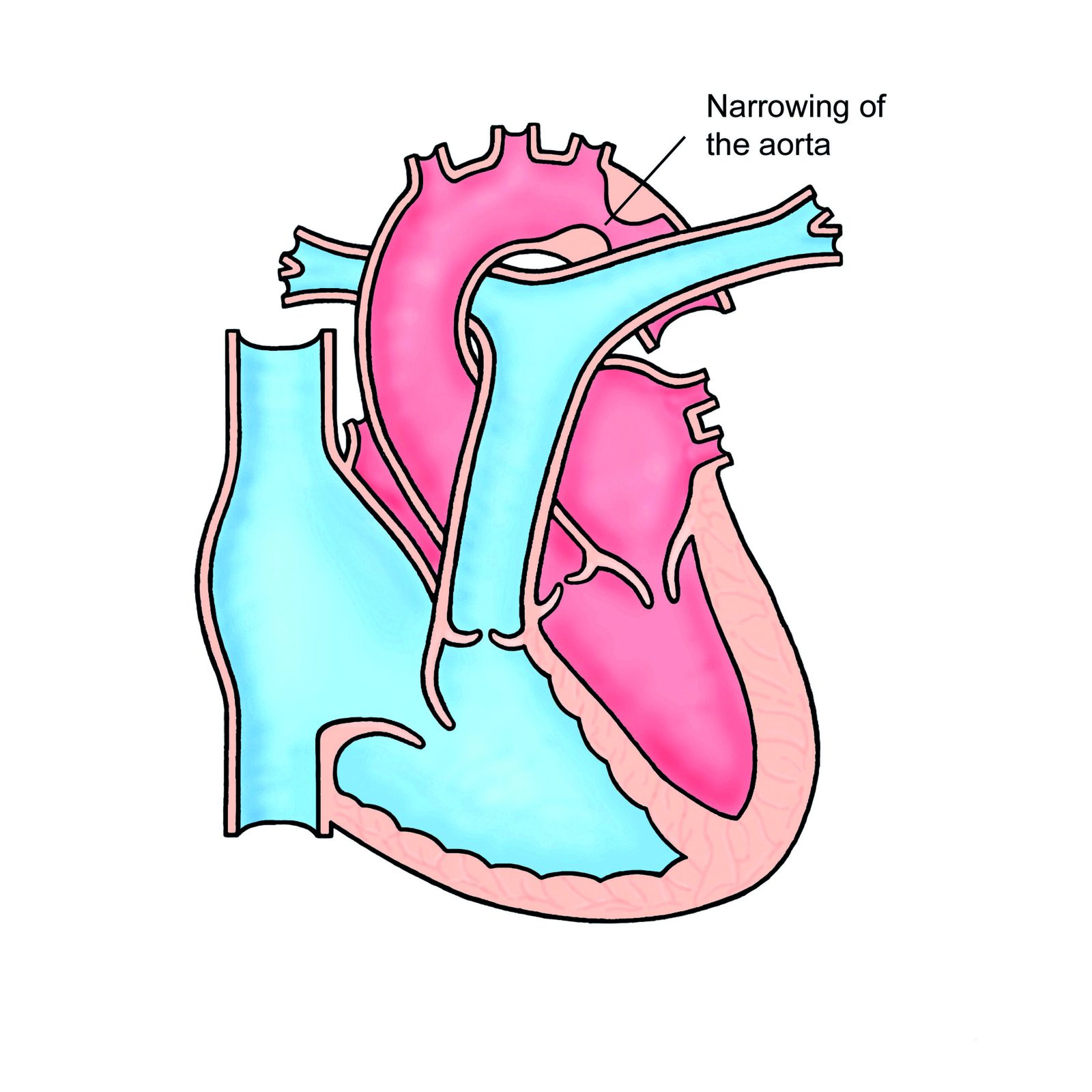
Coarctation of the aorta is a narrowing of the aorta, the main artery leaving the heart, usually just beyond the point where the artery to the left arm branches off. It is present from birth (congenital), but milder forms may not be discovered until adolescence or adulthood, often during investigation of high blood pressure.
Because blood has to be pushed through a narrow section, blood pressure is high in the arms and head and lower in the legs. Over time, untreated coarctation strains the heart and blood vessels, increasing the risk of heart failure, stroke, aneurysms and early heart disease. It can be treated with a stent or surgery, but people need lifelong follow-up even after repair.
When to get urgent help
Seek emergency care for sudden severe chest or back pain, sudden severe headache, stroke symptoms or fainting.
Causes and risk factors
- Congenital: the narrowing develops before birth
- Often associated with a bicuspid aortic valve (in about half of cases)
- Turner syndrome
- Other congenital heart defects, such as ventricular septal defect
- Re-coarctation after childhood repair
How it develops (pathophysiology)
Normal anatomy
The aorta arches over the heart, giving off arteries to the head and arms, then descends through the chest and abdomen to supply the rest of the body. Before birth, a vessel called the ductus arteriosus connects the pulmonary artery to the aorta near the start of the descending aorta; it closes shortly after birth.
What goes wrong
In coarctation, a ridge of tissue narrows the aorta, usually near the site of the ductus arteriosus. One theory is that ductal tissue extends into the aortic wall and constricts as the duct closes.
- Pressure overload: the heart must pump harder to push blood through the narrowing, raising pressure in the aorta and its branches before the narrowing (to the head and arms).
- Low pressure after the narrowing: blood pressure and flow to the lower body are reduced.
- Collateral circulation: over years, the body develops large bypass vessels, mainly enlarged intercostal arteries between the ribs, which carry blood around the narrowing. These can erode the undersides of ribs (rib notching, seen on X-ray).
- Kidney response: reduced blood flow to the kidneys activates the renin-angiotensin system, raising blood pressure further.
- Vascular changes: the arteries above the narrowing become stiff and thickened, and abnormal vessel wall tissue is often present throughout the aorta, which is why high blood pressure can persist even after repair.
- Heart effects: the left ventricle thickens (hypertrophy), and over time may weaken.
Why the symptoms happen
- High blood pressure in the arms results from the obstruction and kidney response.
- Weak or delayed pulses in the legs, and lower leg blood pressure, reflect reduced flow beyond the narrowing.
- Headaches and nosebleeds can result from high pressure in the upper body.
- Leg cramps or tiredness when walking occur from reduced blood supply.
- Heart murmur arises from turbulent flow through the narrowing or collaterals.
How treatment acts on the mechanism
Balloon angioplasty with stenting widens the narrowed segment from inside. Surgical repair removes the narrowed segment and reconnects the aorta, or uses a patch or graft. Relieving the obstruction lowers upper body pressure and improves flow to the lower body. Blood pressure medicines are often still needed because of persistent vessel stiffness.
Types
- Discrete coarctation: short narrowing
- Long-segment or tubular narrowing
- Native (never repaired) or re-coarctation after previous repair
- Isolated or complex (with other heart defects)
Signs and symptoms
Many adults have no symptoms. Possible symptoms include:
- High blood pressure, sometimes hard to control
- Headaches
- Nosebleeds
- Cold feet or leg cramps when walking
- Tiredness or breathlessness on exercise
- Chest pain
- Symptoms of heart failure in late stages
How it is diagnosed
- Blood pressure in both arms and a leg: higher in the arms
- Feeling the pulses: weak or delayed femoral pulses compared with the radial pulse (radio-femoral delay)
- Echocardiogram
- Chest X-ray: may show rib notching or a "3 sign"
- CT or MRI angiography to measure the narrowing
- Cardiac catheterisation to measure the pressure difference, often during treatment
- Assessment for bicuspid aortic valve and brain aneurysms
Possible complications
- Persistent high blood pressure
- Heart failure
- Stroke and brain aneurysm rupture
- Aortic dissection or aneurysm, including at the repair site
- Endocarditis
- Early coronary artery disease
- Re-coarctation after repair
Goals of treatment
Treatment aims to relieve the narrowing, control blood pressure and prevent long-term complications.
Medicines
- Blood pressure medicines, such as beta-blockers, ACE inhibitors or ARBs
- Statins if indicated for cardiovascular risk
Other treatments
- Balloon angioplasty and stenting, the usual first choice in adults
- Surgical repair for complex anatomy
- Lifelong follow-up with a congenital heart disease specialist
Living with it and prevention
- Control blood pressure
- Have regular follow-up imaging
- Discuss exercise with your specialist; heavy weightlifting may be discouraged
- Maintain good dental hygiene to lower endocarditis risk
- Plan pregnancy with specialist advice
Monitoring and follow-up
Regular reviews include blood pressure checks (including exercise or 24-hour monitoring), echocardiograms and periodic CT or MRI to check the repair site for narrowing or aneurysm.
Outlook
With treatment, most adults do well, but they remain at higher lifelong risk of high blood pressure and cardiovascular disease, so regular follow-up is essential.
Lifelong follow-up with a congenital heart team is recommended, even after a successful repair. Regular blood pressure checks, imaging of the aorta and attention to heart health help catch late problems such as re-narrowing, aneurysm or high blood pressure before they cause harm.
Special considerations
- Pregnancy: higher risk of hypertension and aortic complications; needs specialist care
- Turner syndrome: coarctation is common and should be screened for
- Athletes: activity advice depends on the degree of narrowing and blood pressure