
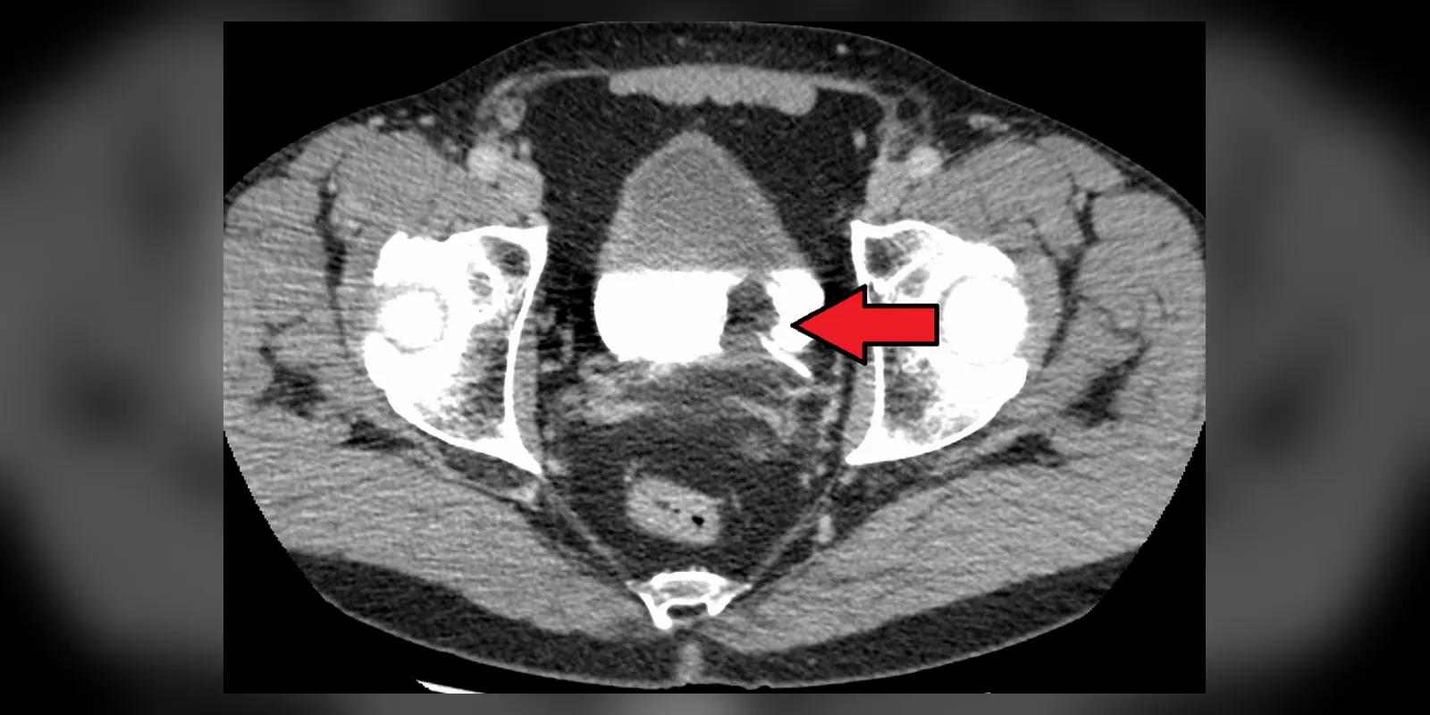
Overview
Bladder cancer is a cancer that begins in the cells lining the inside of the bladder, the organ that stores urine. It is one of the more common cancers, particularly in older people and men, and smoking is its biggest risk factor.
The most common symptom is blood in the urine, which is usually painless and may come and go. Most bladder cancers are found early, when they are confined to the inner lining (non-muscle-invasive), and can be treated by removing the tumour through a telescope inserted into the bladder. However, these cancers often come back, so regular checks are needed. Cancers that have grown into the bladder muscle are more serious and may need removal of the bladder, chemotherapy or radiotherapy.
When to get urgent help
See a doctor promptly for any blood in the urine. Seek urgent care for inability to pass urine, heavy bleeding with clots, or severe pain.
Causes and risk factors
- Smoking: the main cause, responsible for about half of cases
- Workplace chemicals: aromatic amines in dye, rubber, leather, textile and paint industries
- Older age
- Male sex
- Chronic bladder irritation: long-term catheters, bladder stones, recurrent infections
- Schistosomiasis (a parasitic infection), common in parts of Africa and the Middle East
- Previous radiotherapy to the pelvis or chemotherapy with cyclophosphamide
- Arsenic in drinking water
- Family history and genetic conditions, such as Lynch syndrome
How it develops (pathophysiology)
Normal bladder lining
The bladder is lined by urothelium, a specialised layer of cells that stretches as the bladder fills and protects underlying tissue from urine. Beneath it lie connective tissue (lamina propria) and the thick bladder muscle (detrusor).
What goes wrong
- Carcinogen exposure: chemicals from tobacco smoke (such as aromatic amines and polycyclic hydrocarbons) are absorbed into the blood, processed by the liver, and excreted in urine. Concentrated in urine stored in the bladder, they come into prolonged contact with the urothelium, damaging DNA.
- Genetic changes: two main pathways lead to cancer: - Low-grade, non-invasive tumours often have mutations in FGFR3 and HRAS, forming finger-like (papillary) growths that tend to recur but rarely invade. - High-grade, invasive tumours often have mutations in TP53 and RB1, and can develop from flat carcinoma in situ (CIS), with a high risk of invading the muscle.
- Field change: carcinogens affect the whole bladder lining, explaining why tumours often recur in other parts of the bladder.
- Invasion and spread: high-grade cancers invade the bladder muscle, then spread to nearby lymph nodes, organs, bones, lungs and liver.
Chronic inflammation, as in schistosomiasis, can cause squamous cell carcinoma of the bladder.
Why the symptoms happen
- Blood in the urine comes from fragile tumour blood vessels.
- Frequent, urgent or painful urination may result from tumour irritation of the bladder, particularly with CIS.
- Pelvic or back pain suggests spread or blockage of the ureters.
- Weight loss and tiredness occur with advanced cancer.
How treatment acts on the mechanism
Transurethral resection of bladder tumour (TURBT) removes the tumour via the urethra. Intravesical treatments placed in the bladder target the whole lining: BCG (a weakened tuberculosis bacterium) stimulates an immune response that kills cancer cells, and chemotherapy (such as mitomycin or gemcitabine) kills abnormal cells. Cystectomy removes the bladder when cancer invades muscle or high-risk disease does not respond. Chemotherapy before surgery improves survival. Radiotherapy with chemotherapy can preserve the bladder. Immunotherapy and targeted treatments, such as FGFR inhibitors and enfortumab vedotin, treat advanced disease.
Types
- Urothelial carcinoma: about 90 percent
- Squamous cell carcinoma
- Adenocarcinoma
- Non-muscle-invasive bladder cancer: about 75 percent of new cases
- Muscle-invasive bladder cancer
- Carcinoma in situ: flat, high-grade, non-invasive
Signs and symptoms
- Blood in the urine, often painless; urine may look pink, red or brown, or blood may only be found on a urine test
- Needing to urinate more often
- Urgency
- Pain or burning when urinating
- Pelvic or lower back pain
- Swelling of the legs
- Weight loss and tiredness (in advanced cancer)
How it is diagnosed
- Urine tests: for blood, infection and abnormal cells (cytology)
- Cystoscopy: a thin camera passed into the bladder to look at the lining
- Biopsy or TURBT to confirm and stage the cancer
- CT urogram to check the kidneys and ureters
- CT or MRI of the chest, abdomen and pelvis for staging
Possible complications
- Recurrence
- Spread to lymph nodes and other organs
- Blockage of the ureters and kidney damage
- Anaemia from bleeding
- Side effects of treatment, including bladder irritation and, after cystectomy, the need for a urine diversion
Goals of treatment
Treatment aims to remove the cancer, prevent recurrence and progression, preserve bladder function where possible, and control advanced disease.
Medicines
- Intravesical BCG for high-risk non-muscle-invasive cancer
- Intravesical chemotherapy (mitomycin, gemcitabine)
- Neoadjuvant chemotherapy before cystectomy
- Chemotherapy for advanced cancer
- Immunotherapy: pembrolizumab, nivolumab, avelumab
- Antibody-drug conjugates: enfortumab vedotin
- Targeted therapy: erdafitinib for FGFR alterations
Other treatments
- TURBT
- Radical cystectomy with urinary diversion (ileal conduit or new bladder)
- Radiotherapy with chemotherapy (bladder preservation)
- Palliative care
Living with it and prevention
- Stop smoking
- Follow workplace safety rules for chemicals
- Drink plenty of fluids
- Seek prompt investigation of blood in the urine
Monitoring and follow-up
Non-muscle-invasive bladder cancer needs regular cystoscopy, often every three to six months at first, because of the high recurrence rate. After cystectomy, scans monitor for recurrence.
Outlook
Non-muscle-invasive bladder cancer has a good outlook, though recurrence is common. Muscle-invasive cancer is more serious, but can be cured in many people with surgery or chemoradiotherapy. Advanced cancer has a poorer outlook, though newer treatments are improving survival.
Special considerations
- Smokers and ex-smokers: highest risk
- Women: often diagnosed later because blood in urine may be attributed to infection
- People from areas with schistosomiasis: higher risk of squamous cell carcinoma