
Overview
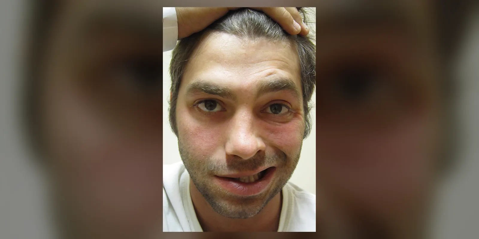
Bell's palsy is sudden weakness or paralysis of the muscles on one side of the face, caused by inflammation of the facial nerve. It usually develops over hours to a couple of days and can make one side of the face droop, affecting the ability to smile, close the eye, raise the eyebrow and eat or drink normally.
Bell's palsy is the most common cause of facial paralysis. It can happen at any age but is most common between 15 and 60, and in pregnancy and people with diabetes. Although it can be frightening and is often mistaken for a stroke, most people recover fully within weeks to months, especially when treated early with steroids. Protecting the eye is important while it cannot close fully.
When to get urgent help
Call emergency services if facial weakness comes with weakness or numbness of the arm or leg, slurred speech, confusion, severe headache or vision problems, which may indicate a stroke. Seek prompt care for a painful red eye or loss of vision.
Causes and risk factors
The exact cause is unknown, but it is thought to be linked to:
- Viral infections, especially reactivation of herpes simplex virus (the cold sore virus), and sometimes varicella zoster, Epstein-Barr virus and others
- Immune or inflammatory reactions affecting the facial nerve
Risk factors include:
- Pregnancy, especially the third trimester
- Diabetes
- Upper respiratory infections
- High blood pressure
- Obesity
- Family history
How it develops (pathophysiology)
Normal facial nerve function
The facial nerve (seventh cranial nerve) leaves the brainstem, passes through a narrow bony canal in the skull (the facial canal in the temporal bone), and exits just below the ear. It controls:
- Facial expression muscles, such as those for smiling, frowning, closing the eyes and raising the eyebrows
- Taste from the front two thirds of the tongue
- Tear and saliva production
- A small muscle in the ear (stapedius) that dampens loud sounds
What goes wrong
- Inflammation: a viral infection or immune reaction causes inflammation of the facial nerve.
- Swelling in a tight space: the nerve swells, but it is enclosed in a narrow bony canal, especially at its narrowest part (the labyrinthine segment).
- Compression and reduced blood flow: swelling compresses the nerve and its blood supply, interrupting nerve signals.
- Nerve damage: if compression is mild, nerve conduction is temporarily blocked (neurapraxia), and recovery is rapid. If severe, nerve fibres degenerate (axonotmesis), and recovery depends on regrowth, which is slower and may be incomplete.
- Recovery and aberrant regrowth: as nerve fibres regrow, some may connect to the wrong muscles (synkinesis), causing unwanted movements, such as the eye closing when smiling, or tears when eating (crocodile tears).
Why the symptoms happen
- Drooping of one side of the face and inability to smile reflect loss of nerve signals to facial muscles.
- Inability to close the eye fully occurs because the orbicularis oculi muscle is weak.
- Dry eye or watery eye occurs from reduced blinking, altered tear production and poor tear drainage.
- Drooling and difficulty eating reflect weak mouth muscles.
- Altered taste results from involvement of taste fibres.
- Sensitivity to loud sounds (hyperacusis) results from weakness of the stapedius muscle.
- Pain behind the ear often occurs early, from inflammation.
Importantly, because the problem is in the nerve itself, the forehead is affected, unlike in a stroke, where the forehead is often spared.
How treatment acts on the mechanism
Corticosteroids (prednisolone), started within 72 hours, reduce inflammation and swelling of the nerve, improving the chance of full recovery. Antivirals may be added in severe cases, though benefit is uncertain. Eye protection with lubricating drops, ointment and taping prevents corneal damage. Physiotherapy and facial exercises support recovery and reduce synkinesis.
Types
- Mild (incomplete) Bell's palsy: some movement remains; usually recovers fully
- Complete Bell's palsy: total paralysis on one side; recovery may be slower
- Graded using scales such as the House-Brackmann scale
Signs and symptoms
- Sudden weakness or drooping of one side of the face
- Difficulty smiling, closing the eye or raising the eyebrow on one side
- Drooling
- Difficulty eating and drinking
- Dry or watery eye
- Pain behind or around the ear
- Altered taste
- Sensitivity to loud sounds
- Symptoms usually develop over 24 to 72 hours
How it is diagnosed
- Clinical examination: weakness of the whole of one side of the face, including the forehead
- Neurological examination to exclude stroke and other causes
- Ear examination to exclude infection or shingles (Ramsay Hunt syndrome)
- Blood tests for diabetes, Lyme disease or other causes when relevant
- Imaging (MRI) if the presentation is atypical, recovery does not occur, or other nerve problems are present
- Nerve conduction tests in severe cases
Possible complications
- Incomplete recovery and permanent facial weakness
- Synkinesis: involuntary facial movements
- Crocodile tears
- Corneal damage from inability to close the eye
- Psychological distress
Goals of treatment
Treatment aims to speed recovery, protect the eye, reduce complications and support emotional wellbeing.
Medicines
- Prednisolone, started within 72 hours of onset
- Antivirals (such as aciclovir or valaciclovir) with steroids in severe cases
- Lubricating eye drops and ointment
Other treatments
- Eye protection: wearing glasses, taping the eye closed at night
- Facial physiotherapy and exercises
- Botulinum toxin injections for synkinesis
- Surgery in rare cases of persistent paralysis, to improve eye closure or facial symmetry
Living with it and prevention
Bell's palsy cannot be prevented. During recovery:
- Protect the affected eye
- Do facial exercises as advised
- Chew on the unaffected side
- Seek support if the change in appearance is distressing
Monitoring and follow-up
Most people start to improve within three weeks. If there is no improvement by three months, referral to a specialist is recommended.
Outlook
Most people recover fully, often within three to six months. About 70 to 85 percent recover completely, especially with early treatment. Some have lasting weakness or synkinesis.
Special considerations
- Pregnancy: higher risk, particularly in the third trimester; steroids may still be used
- Children: usually recover well
- People with diabetes: higher risk and possibly slower recovery
- Recurrence: occurs in a small number of people; recurrent or bilateral palsy needs investigation