
Overview
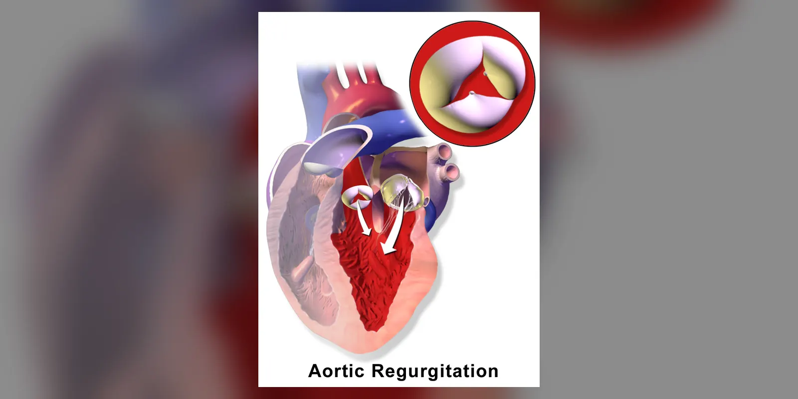
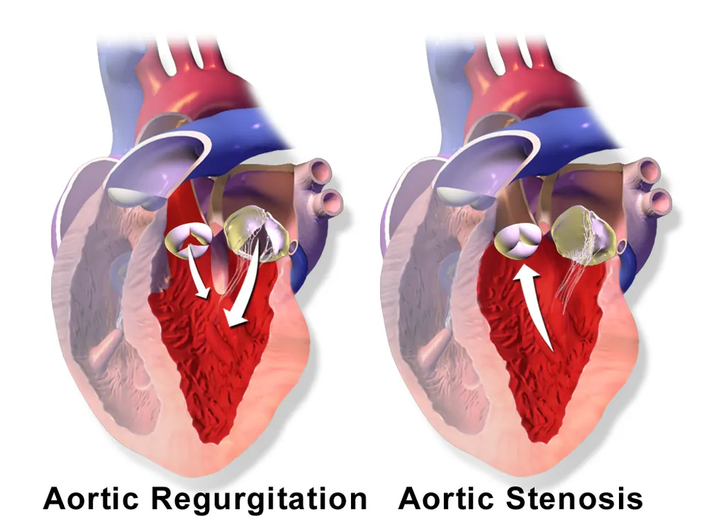
Aortic regurgitation (also called aortic insufficiency) is a condition in which the aortic valve, the valve between the left ventricle and the aorta, does not close properly. With each heartbeat, some blood leaks back into the heart instead of flowing to the body.
The heart compensates by enlarging and pumping more forcefully, so mild to moderate regurgitation may cause no symptoms for many years. Over time, the extra work can weaken the heart, leading to breathlessness, tiredness and heart failure. Aortic regurgitation can also develop suddenly, for example from infection of the valve or aortic dissection, which is a medical emergency. Treatment ranges from monitoring to valve replacement surgery.
Because symptoms often appear late, regular check-ups with heart scans are important for people known to have a leaky aortic valve, so that surgery can be offered before the heart muscle is permanently weakened.
When to get urgent help
Seek emergency care for sudden severe breathlessness, chest pain, fainting, or sudden severe tearing chest or back pain.
Causes and risk factors
Valve leaflet problems:
- Bicuspid aortic valve (born with two leaflets instead of three)
- Rheumatic heart disease
- Infective endocarditis
- Age-related degeneration
Aortic root widening:
- High blood pressure
- Marfan syndrome and other connective tissue disorders
- Aortic aneurysm
- Aortic dissection (sudden)
- Inflammatory conditions: ankylosing spondylitis, syphilis, giant cell arteritis
How it develops (pathophysiology)
Normal valve function
The aortic valve has three thin leaflets attached to a ring at the base of the aorta (the aortic root). When the left ventricle contracts (systole), the leaflets open to let blood into the aorta. When the ventricle relaxes (diastole), pressure in the aorta pushes the leaflets together, sealing the valve.
What goes wrong
Regurgitation occurs when the leaflets are damaged or when the aortic root widens so the leaflets cannot meet.
Chronic aortic regurgitation:
- During diastole, blood leaks back from the aorta into the left ventricle.
- The ventricle receives blood from both the left atrium and the leak, causing volume overload.
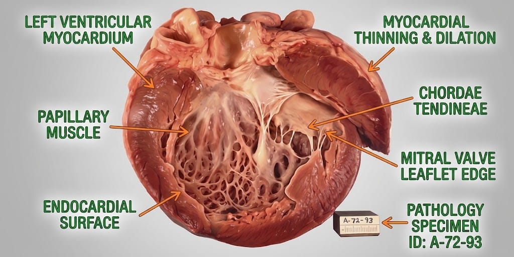
- The ventricle adapts by enlarging (eccentric hypertrophy), allowing it to eject a larger volume with each beat. The amount reaching the body stays near normal.
- The large stroke volume ejected into the aorta raises systolic pressure, while the leak lowers diastolic pressure, producing a wide pulse pressure.
- After years, the ventricle cannot keep enlarging. Its muscle stretches beyond its ability to contract efficiently, pressure inside rises, and heart failure develops. Damage may become irreversible if surgery is delayed.
Acute aortic regurgitation:
The ventricle has no time to enlarge. A sudden large leak causes a rapid rise in pressure in the ventricle and lungs, leading to fluid on the lungs (pulmonary oedema) and shock.
Why the symptoms happen
- Breathlessness and tiredness occur as the heart fails and pressure rises in the lungs.
- Pounding heartbeat, especially lying on the left side, reflects the large stroke volume.
- Chest pain may result from reduced coronary blood flow, because coronary arteries fill during diastole when aortic pressure is low.
- Visible pulsations (such as head bobbing or bounding pulses) reflect the wide pulse pressure.
- Murmur: a soft, high-pitched sound heard after the second heart sound, as blood flows backwards.
How treatment acts on the mechanism
Blood pressure medicines (vasodilators such as ACE inhibitors or calcium channel blockers) reduce resistance in the aorta, encouraging forward flow. Surgical valve replacement or repair stops the leak, relieving the volume load. Surgery is timed before the ventricle becomes permanently damaged, guided by symptoms and measurements of ventricle size and function. Aortic root replacement is needed when the root is enlarged.
Types
- Acute aortic regurgitation: sudden, often from endocarditis or dissection
- Chronic aortic regurgitation: develops slowly
- Graded mild, moderate or severe on echocardiography
Signs and symptoms
- Often no symptoms for years
- Breathlessness on exertion, later at rest or lying flat
- Tiredness and reduced exercise tolerance
- Palpitations or a pounding heartbeat
- Chest pain
- Dizziness or fainting
- Swollen ankles in heart failure
Acute: sudden severe breathlessness, low blood pressure and collapse.
How it is diagnosed
- Listening to the heart: a diastolic murmur
- Blood pressure: wide pulse pressure
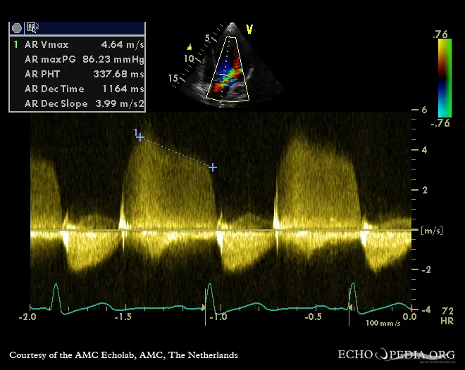
- Echocardiogram, the main test for severity and heart size
- ECG
- Cardiac MRI to measure the leak and heart function
- CT to assess the aortic root
- Blood tests and blood cultures if endocarditis is suspected
Possible complications
- Heart failure
- Arrhythmias
- Infective endocarditis
- Sudden death (rare)
- Complications of an enlarged aortic root, such as dissection
Goals of treatment
Treatment aims to relieve symptoms, protect heart function, and replace or repair the valve at the right time.
Medicines
- Blood pressure control with ACE inhibitors, ARBs or calcium channel blockers
- Diuretics for fluid overload
- Beta-blockers or ARBs in Marfan syndrome to slow aortic root enlargement
- Antibiotics for endocarditis
Other treatments
- Aortic valve replacement (mechanical or biological valve)
- Valve repair in selected cases
- Aortic root replacement, sometimes sparing the valve
- Transcatheter valve replacement in selected high-risk patients
- Emergency surgery for acute severe regurgitation
Living with it and prevention
- Control blood pressure
- Maintain good dental health and seek treatment for infections
- Attend regular echocardiograms
- Stay active within advice
- People with Marfan syndrome need specialist follow-up
Monitoring and follow-up
People with moderate or severe regurgitation have regular echocardiograms, often every 6 to 12 months, to monitor the size and function of the left ventricle.
Outlook
Mild regurgitation may never cause problems. Severe regurgitation leads to heart failure without treatment, but timely valve surgery offers good long-term outcomes.
Special considerations
- Pregnancy: usually well tolerated in chronic regurgitation but needs monitoring
- Bicuspid valve or Marfan syndrome: family members may need screening
- Athletes: exercise advice depends on severity