
Overview
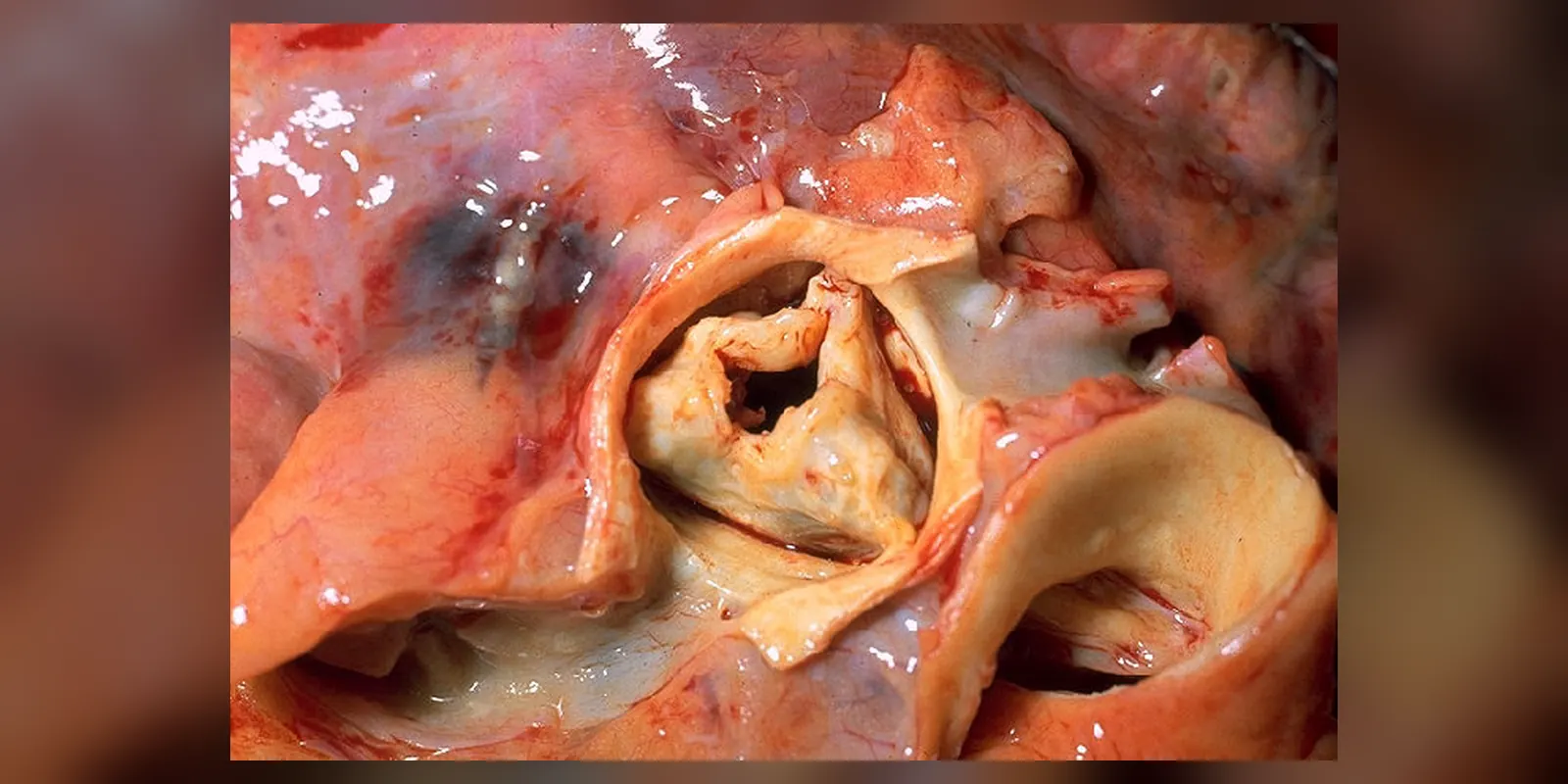
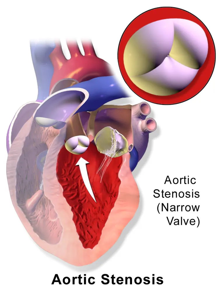
Aortic stenosis is a narrowing of the aortic valve, which controls blood flow from the heart's main pumping chamber (the left ventricle) into the aorta and the rest of the body. When the valve becomes stiff and narrow, the heart has to work harder to push blood through it.
It is the most common serious valve disease in older people in high-income countries, usually caused by gradual build-up of calcium on the valve. It may cause no symptoms for years, but once symptoms such as chest pain, breathlessness or fainting develop, the outlook without treatment is poor. Valve replacement, either by open-heart surgery or by a less invasive procedure called TAVI, is highly effective.
When to get urgent help
Seek urgent care for fainting, chest pain at rest, sudden severe breathlessness, or palpitations with dizziness.
Causes and risk factors
- Age-related calcific degeneration: the most common cause in older adults
- Bicuspid aortic valve: present from birth, leading to earlier stenosis, often in people aged 50 to 70
- Rheumatic heart disease, more common in low- and middle-income countries
- Risk factors: older age, high blood pressure, high cholesterol, smoking, diabetes, chronic kidney disease, and high lipoprotein(a)
How it develops (pathophysiology)
Normal valve function
The aortic valve has three thin, flexible leaflets that open fully when the left ventricle contracts and close when it relaxes. Normally the valve opening is about 3 to 4 square centimetres, allowing blood to flow out freely with almost no pressure difference across it.
What goes wrong
In calcific aortic stenosis, the process resembles atherosclerosis:
- Endothelial injury: mechanical stress on the leaflets, especially where they flex, damages the lining.
- Lipid and inflammation: LDL cholesterol and lipoprotein(a) enter the leaflet, become oxidised and attract inflammatory cells.
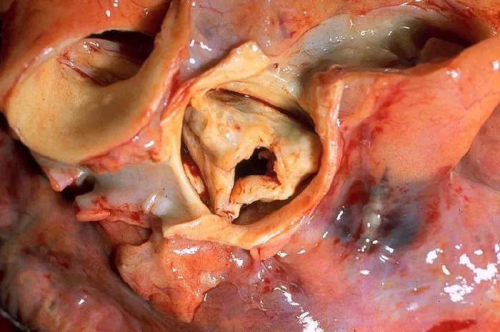
- Calcification: valve cells (interstitial cells) change into bone-forming cells, laying down calcium. Leaflets thicken and stiffen.
- Narrowing: as calcium builds up over years, the leaflets cannot open fully, and the valve area shrinks. Severe stenosis is generally an area below about 1 square centimetre.
A bicuspid valve experiences higher mechanical stress, so it calcifies earlier.
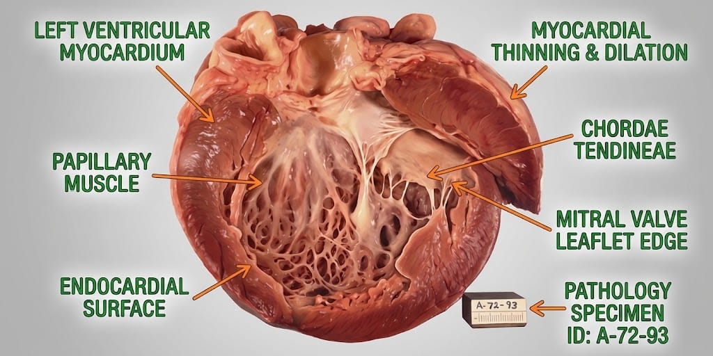
Effects on the heart
- The left ventricle must generate much higher pressure to push blood through the narrow valve (pressure overload).
- It adapts by thickening its walls (concentric hypertrophy), which maintains output for years.
- The thick muscle becomes stiff and needs more oxygen, but blood flow through the coronary arteries does not increase enough. This can cause ischaemia even without coronary artery disease.
- Eventually, the muscle becomes scarred (fibrosis), the ventricle fails, and pressure builds up in the lungs.
Why the symptoms happen
- Chest pain (angina) occurs because the thickened muscle needs more oxygen than it receives.
- Breathlessness results from the stiff ventricle and raised pressure in the lungs.
- Fainting or dizziness on exertion occurs because the narrow valve prevents cardiac output from rising when blood vessels in the muscles widen during exercise, so blood pressure drops.
- Tiredness and reduced exercise capacity reflect limited output.
- Ejection systolic murmur arises from turbulent flow through the narrowed valve.
How treatment acts on the mechanism
No medicine can reverse calcification. Aortic valve replacement, either surgical (SAVR) or transcatheter (TAVI), removes the obstruction. TAVI places a new valve inside the old one via a catheter, usually through the groin artery, without open-heart surgery. Relieving the obstruction lowers pressure in the ventricle, allowing symptoms to improve and the thickened muscle to partly recover.
Types
- Graded mild, moderate or severe on echocardiography
- Severe high-gradient stenosis
- Low-flow, low-gradient stenosis, often with weak heart muscle
- Calcific, bicuspid or rheumatic
Signs and symptoms
Often no symptoms until stenosis is severe. Then:
- Chest pain or tightness on exertion
- Breathlessness on exertion, later at rest
- Dizziness or fainting, especially during exercise
- Tiredness and reduced ability to exercise
- Palpitations
- Swollen ankles in heart failure
How it is diagnosed
- Listening to the heart: a harsh murmur radiating to the neck
- Echocardiogram, the main test, measuring valve area, flow speed and pressure gradient
- ECG
- CT scan to measure calcium and plan TAVI
- Exercise testing in people without clear symptoms
- Coronary angiography before valve replacement
- Blood tests, such as BNP
Possible complications
- Heart failure
- Sudden cardiac death
- Arrhythmias, including atrial fibrillation
- Bleeding from the gut (Heyde's syndrome)
- Endocarditis
- Stroke
Goals of treatment
The goals are to monitor until intervention is needed, then replace the valve to relieve symptoms, prevent heart failure and improve survival.
Medicines
- No medicine slows stenosis
- Diuretics carefully for fluid overload
- Blood pressure control, avoiding sudden drops
- Treatment of atrial fibrillation and other conditions
Other treatments
- Surgical aortic valve replacement (SAVR)
- Transcatheter aortic valve implantation (TAVI)
- Balloon valvuloplasty as a temporary bridge in selected patients
- Regular monitoring for asymptomatic people
Living with it and prevention
- Report new symptoms promptly
- Avoid very strenuous exercise if stenosis is severe
- Keep blood pressure and cholesterol controlled
- Do not smoke
- Maintain good dental health to reduce endocarditis risk
Monitoring and follow-up
Echocardiograms are repeated every one to three years for mild to moderate stenosis and every six to twelve months for severe stenosis. After valve replacement, regular follow-up checks valve function.
Outlook
Without treatment, severe symptomatic aortic stenosis has a poor outlook, with high mortality within a few years. Valve replacement restores near-normal life expectancy for many people.
Special considerations
- Older and frail people: TAVI offers an effective option
- Bicuspid valve: earlier onset; family members may be screened
- Pregnancy: severe stenosis is high-risk and needs specialist planning