
Overview
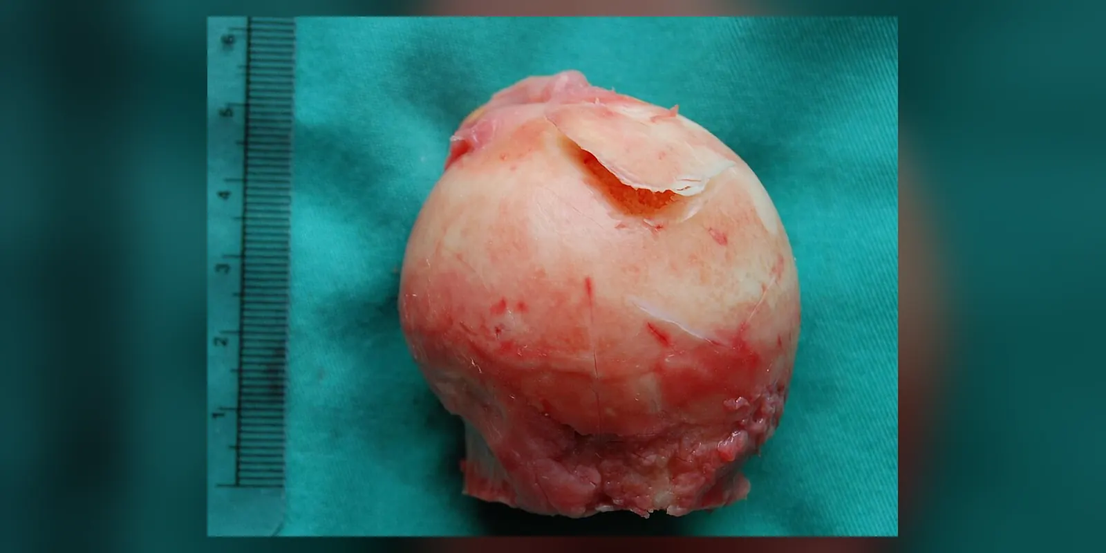
Avascular necrosis, also called osteonecrosis, is the death of bone tissue caused by a lack of blood supply. Without blood, bone cells die, and over time the affected bone can weaken and collapse, damaging the nearby joint.
It most often affects the head of the femur (thigh bone) at the hip, but can also affect the shoulder, knee, ankle and other bones. It commonly occurs in people aged 30 to 50. Causes include long-term high-dose steroid use, heavy alcohol use, hip fractures and dislocations, sickle cell disease and some other medical conditions. Early stages may cause no symptoms, while later stages cause pain and stiffness. Treatment depends on the stage and ranges from medicines and protected weight-bearing to surgery, including joint replacement.
When to get urgent help
Seek urgent care for severe sudden hip or joint pain, inability to bear weight, or joint pain with fever.
Causes and risk factors
- Long-term high-dose corticosteroids: one of the most common causes
- Heavy alcohol use
- Injury: hip fractures, dislocations
- Sickle cell disease and other blood disorders
- Radiation therapy
- Chemotherapy
- Organ transplantation, particularly kidney
- Lupus and other autoimmune diseases
- HIV infection
- Gaucher disease
- Decompression sickness in divers
- Bisphosphonates and denosumab can cause osteonecrosis of the jaw
- Idiopathic: no cause found in some people
How it develops (pathophysiology)
Normal bone blood supply
Bone is living tissue with a rich blood supply delivering oxygen and nutrients to bone cells (osteocytes) and bone marrow. Some areas, such as the femoral head, rely on a few end arteries with limited back-up supply. If these vessels are damaged, the bone they supply has no alternative blood source.
What goes wrong
Blood supply can be interrupted in several ways:
- Mechanical disruption: fractures or dislocations tear blood vessels supplying the femoral head.
- Fat emboli and increased marrow pressure: steroids and alcohol enlarge fat cells in the bone marrow and raise lipid levels. Swollen fat cells raise pressure inside the bone, compressing small blood vessels, and fat droplets may block vessels.
- Blood vessel blockage: sickle-shaped red cells, clotting disorders or nitrogen bubbles (in decompression sickness) block small vessels.
- Vessel damage: radiation and some diseases damage vessel walls.
Stages of damage
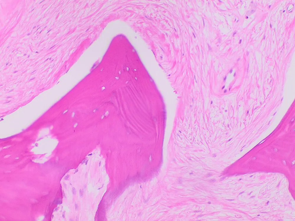
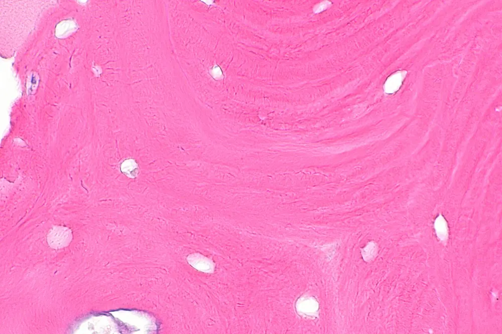
- Ischaemia: without blood, bone cells and marrow die within 6 to 24 hours.
- Repair attempt: the surrounding living bone tries to repair the dead area, with new blood vessels growing in and bone being removed and rebuilt. Removal of dead bone may outpace new bone formation, weakening the area.
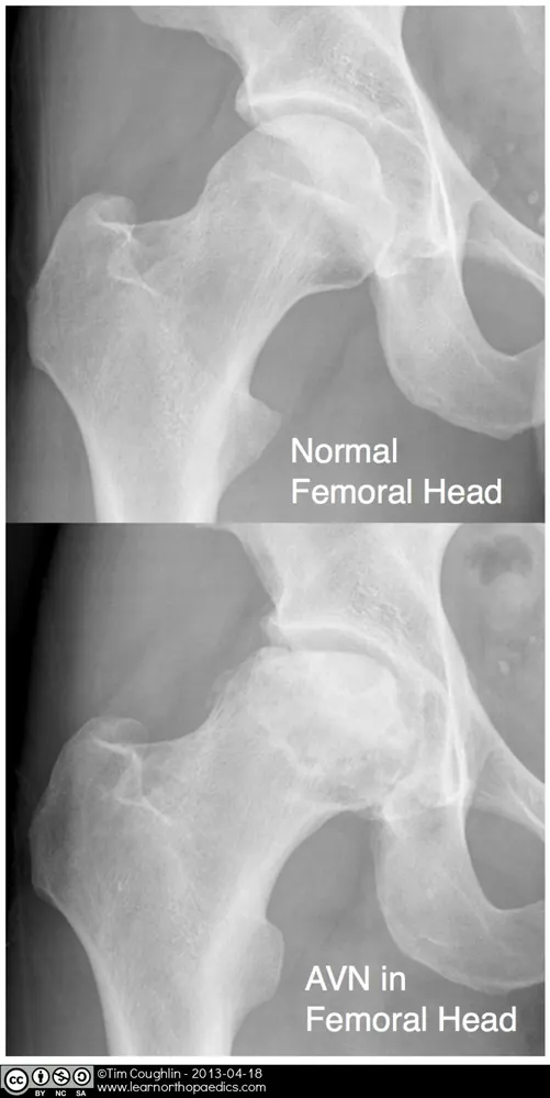
- Subchondral fracture: weakened bone just beneath the joint cartilage develops tiny fractures, sometimes seen on X-ray as a "crescent sign".
- Collapse: the joint surface flattens and collapses.
- Secondary arthritis: the irregular joint surface damages cartilage, causing osteoarthritis.
Why the symptoms happen
- No symptoms early because dead bone itself does not cause pain until it starts to fail.
- Groin or hip pain (or pain in the affected joint) occurs with raised pressure in the bone, microfractures and collapse.
- Pain on weight-bearing, later at rest and at night.
- Stiffness and reduced movement result from joint damage and arthritis.
- Limping results from pain and joint deformity.
How treatment acts on the mechanism
Reducing risk factors (such as steroids and alcohol) may prevent progression. Protected weight-bearing reduces stress on weakened bone. Core decompression drills into the bone to relieve pressure and stimulate new blood vessel growth, sometimes with bone grafts or bone marrow cells. Osteotomy shifts weight away from the damaged area. Joint replacement replaces the collapsed joint and relieves pain.
Types
- By site: femoral head (most common), humeral head, knee (femoral condyles), talus, scaphoid, jaw
- Traumatic or non-traumatic
- Staged by imaging (for example Ficat or ARCO staging), from early changes seen only on MRI to collapse and arthritis
Signs and symptoms
- No symptoms in early stages
- Pain in the groin, thigh or buttock (hip), or in the affected joint
- Pain on weight-bearing, later at rest or at night
- Stiffness and reduced range of movement
- Limping
- Pain may affect both hips
How it is diagnosed
- History of risk factors such as steroid use or alcohol
- X-rays: normal early; later show bone changes, collapse and arthritis
- MRI: the most sensitive test for early disease
- CT or bone scan in some cases
- Blood tests for underlying conditions
Possible complications
- Collapse of the bone
- Osteoarthritis
- Disability and chronic pain
- Involvement of the other joint
Goals of treatment
Treatment aims to relieve pain, preserve the joint where possible, slow progression and restore function.
Medicines
- Pain relief: paracetamol, NSAIDs
- Bisphosphonates in some early cases, though evidence is mixed
- Treatment of underlying conditions
- Reducing or stopping steroids if possible
Other treatments
- Protected weight-bearing with crutches
- Physiotherapy to maintain movement and strength
- Core decompression, with or without bone grafts or cell therapy
- Vascularised bone grafts
- Osteotomy
- Joint replacement for advanced collapse or arthritis
Living with it and prevention
- Use steroids at the lowest effective dose for the shortest time
- Limit alcohol
- Follow safe diving practices
- Manage sickle cell disease and other conditions
- Have dental checks before starting bisphosphonates or denosumab
Monitoring and follow-up
Imaging is repeated to monitor progression. People with one affected hip are monitored for the other side.
Outlook
Early-stage disease may stabilise with treatment, but many cases progress to collapse. Joint replacement provides good pain relief and function.
Special considerations
- Children: Perthes disease is a form of avascular necrosis of the hip in children
- People with sickle cell disease: high risk, often both hips
- Transplant recipients and people on long-term steroids: should report joint pain early