
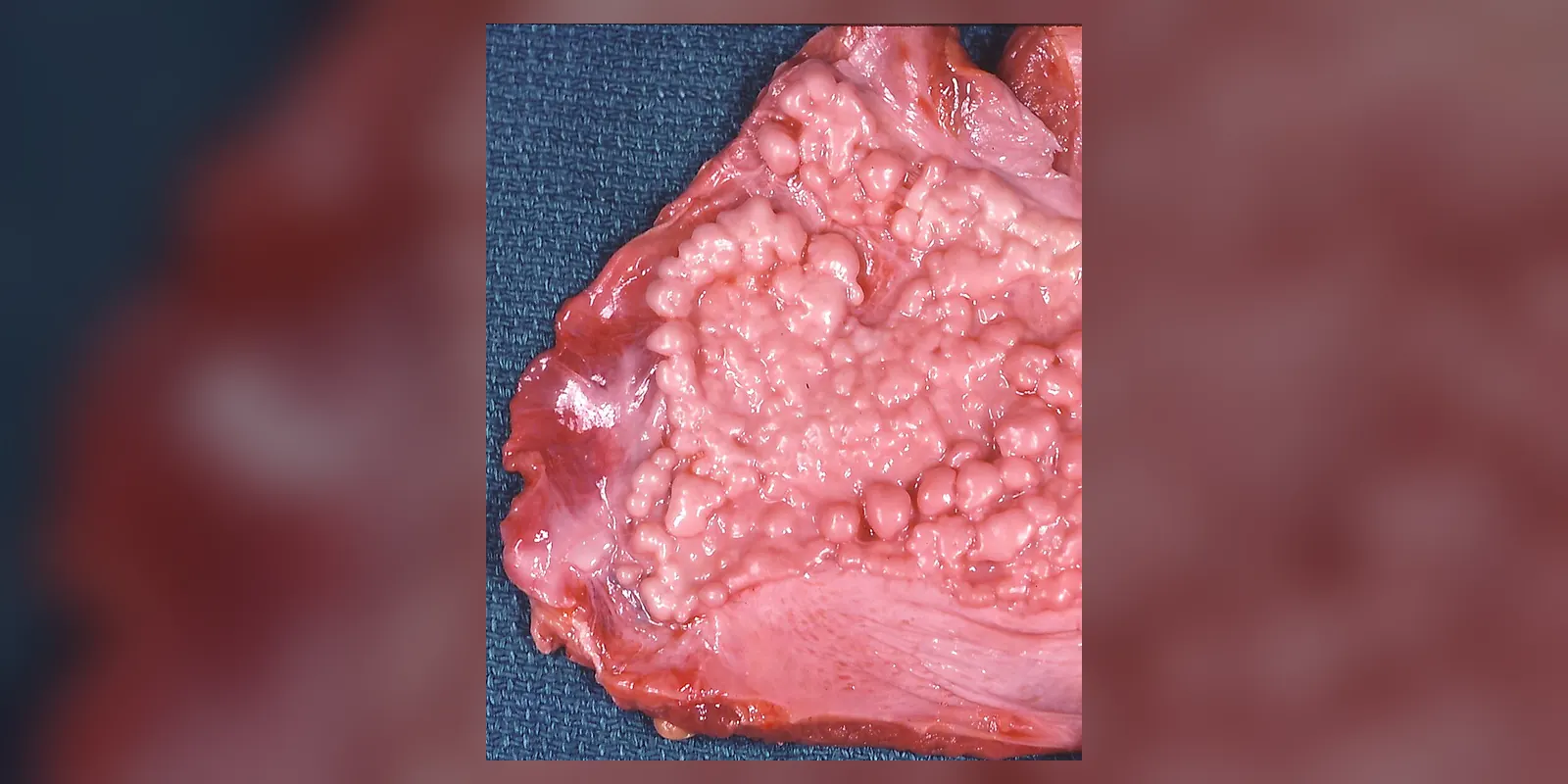
Overview
Asbestosis is a long-term lung disease caused by breathing in asbestos fibres over many years. The fibres lodge deep in the lungs and cause scarring (fibrosis), which makes the lungs stiff and reduces their ability to transfer oxygen into the blood.
It usually develops 20 to 40 years after exposure, mainly in people who worked in construction, shipbuilding, insulation, demolition and manufacturing before asbestos was banned. Asbestosis causes increasing breathlessness and a dry cough and cannot be reversed. Asbestos exposure also increases the risk of lung cancer and mesothelioma, a cancer of the lung lining. Stopping smoking, supportive treatment and pulmonary rehabilitation help people manage symptoms.
Although asbestos is now banned in many countries, it remains in many older buildings, and people who worked with it decades ago are still being diagnosed. Asbestosis is a recognised industrial disease, and affected people may be entitled to compensation or benefits.
When to get urgent help
Seek urgent care for rapidly worsening breathlessness, coughing up blood, chest pain, or signs of a chest infection with fever.
Causes and risk factors
- Long-term inhalation of asbestos fibres, usually at work
- High-risk occupations (especially before the 1980s and 1990s): shipbuilding, construction, insulation fitting, plumbing, electrical work, demolition, asbestos mining and manufacturing, brake and clutch repair
- Secondary exposure: family members washing contaminated work clothes
- Risk increases with the amount and duration of exposure
- Smoking greatly increases the risk of lung cancer and may worsen asbestosis
How it develops (pathophysiology)
Normal lung function
The lungs contain millions of tiny air sacs (alveoli) surrounded by capillaries. The walls between air and blood are extremely thin, allowing oxygen to pass quickly into the blood. Alveolar macrophages, immune cells living in the air sacs, engulf and remove inhaled particles.
What goes wrong
- Fibre inhalation: asbestos fibres are long and thin. Smaller fibres reach the deepest parts of the lungs, especially the lower lobes, and can work their way into the lung tissue and the pleura (lung lining).
- Frustrated phagocytosis: macrophages try to engulf the fibres but cannot fully surround long fibres. They become persistently activated.
- Inflammation and oxidative stress: activated macrophages release reactive oxygen species, inflammatory cytokines (such as TNF-alpha and IL-1) and growth factors, especially transforming growth factor beta (TGF-beta). Iron on fibre surfaces adds to free radical production.
- Fibroblast activation: growth factors stimulate fibroblasts to multiply and produce large amounts of collagen in the walls between alveoli (interstitium).
- Progressive fibrosis: scarring builds up over decades, starting around small airways in the lower lobes and spreading. The lungs become stiff and shrink, and the thin air-blood barrier thickens.
- Persistence: asbestos fibres are not broken down, so inflammation and scarring can continue long after exposure ends.
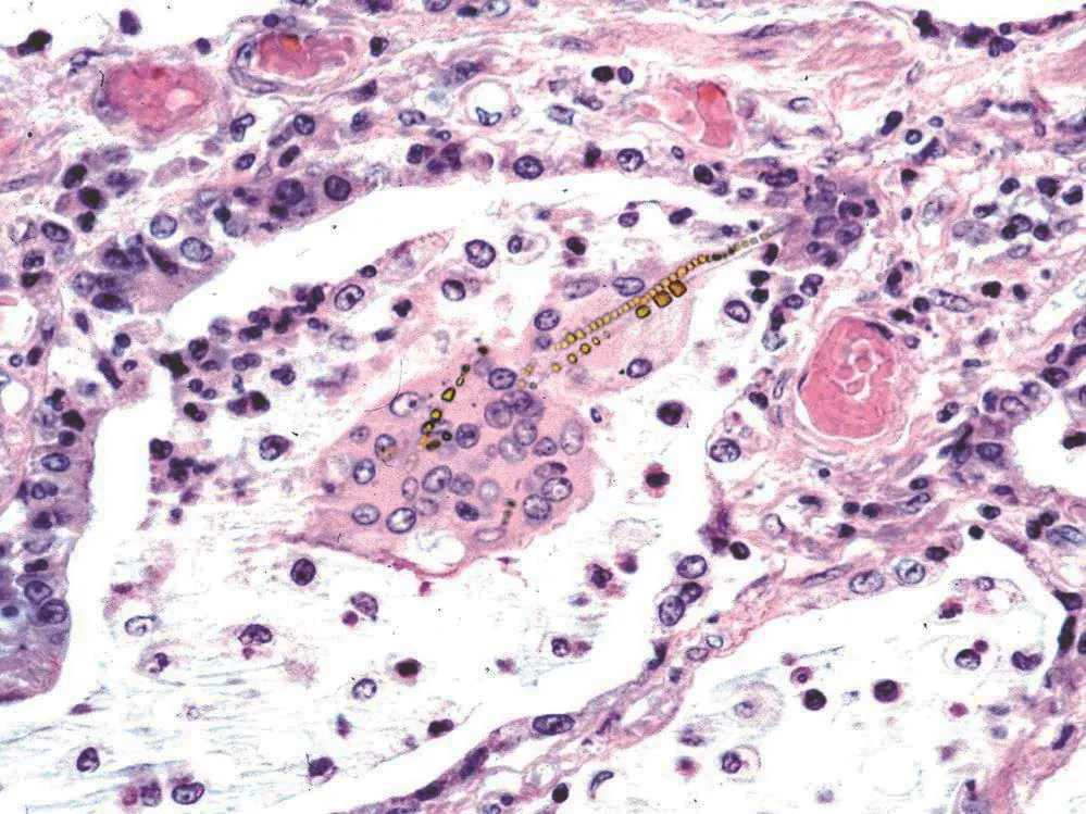
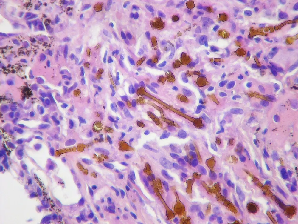
Fibres coated with iron and protein, called asbestos bodies, can be seen under the microscope.
Why the symptoms happen
- Breathlessness, first on exertion, then at rest, results from stiff lungs and impaired oxygen transfer.
- Dry cough reflects irritation and scarring.
- Crackles heard at the lung bases arise from stiff alveoli popping open.
- Finger clubbing may develop in advanced disease.
- Low oxygen levels reflect thickened alveolar walls.
- Right heart strain (cor pulmonale) develops when scarring raises pressure in lung blood vessels.
How treatment acts on the mechanism
There is no treatment that removes asbestos fibres or reverses scarring. Stopping smoking reduces further lung injury and cancer risk. Antifibrotic medicines such as nintedanib may slow fibrosis in progressive disease. Oxygen therapy compensates for impaired gas transfer. Pulmonary rehabilitation improves exercise capacity. Vaccination reduces infections that worsen breathing.
Types
- Asbestosis: fibrosis of lung tissue
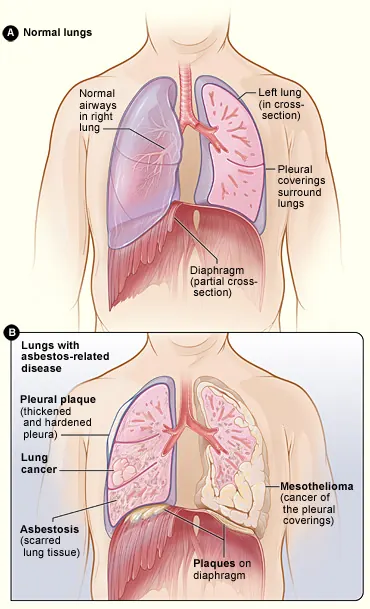
- Related asbestos diseases: pleural plaques, diffuse pleural thickening, benign asbestos pleural effusion, mesothelioma and lung cancer
Signs and symptoms
- Gradually worsening breathlessness
- Persistent dry cough
- Tiredness
- Chest tightness or pain
- Crackling sounds when breathing
- Clubbed fingertips in advanced disease
- Weight loss
Symptoms usually come on slowly, and many people put their breathlessness down to ageing or being unfit. A cough that does not go away and breathlessness climbing stairs are often the first signs.
How it is diagnosed
- Occupational history of asbestos exposure
- Chest X-ray: shadows in the lower lungs and pleural plaques
- High-resolution CT scan: the most sensitive test
- Lung function tests: reduced lung volumes and gas transfer
- Oxygen saturation and exercise testing
- Biopsy rarely needed
Possible complications
- Respiratory failure
- Pulmonary hypertension and right heart failure
- Lung cancer
- Mesothelioma
- Chest infections
Goals of treatment
Treatment aims to relieve symptoms, slow progression, prevent complications and monitor for cancer.
Medicines
- Antifibrotic medicines (such as nintedanib) for progressive fibrosis, in some cases
- Inhalers if airway narrowing is also present
- Antibiotics for chest infections
- Vaccinations against flu, pneumococcus and COVID-19
Other treatments
- Stopping smoking
- Pulmonary rehabilitation
- Oxygen therapy for low oxygen levels
- Lung transplantation in selected cases
- Palliative care for advanced disease
- Compensation claims, which may be available for occupational disease
Living with it and prevention
- Follow strict asbestos safety regulations at work
- Never disturb asbestos-containing materials without professional removal
- Use proper protective equipment
- Stop smoking if previously exposed
If you think your home or workplace contains asbestos, leave it undisturbed and seek advice from a licensed professional before any building work.
Monitoring and follow-up
Regular lung function tests and imaging monitor progression. People with asbestos exposure should report new symptoms such as chest pain, weight loss or coughing blood promptly.
Outlook
Asbestosis usually progresses slowly over years. Outcomes depend on the extent of scarring and whether cancer develops. Many people live for many years after diagnosis.
Special considerations
- Former asbestos workers: should tell their doctor about past exposure
- Family members with secondary exposure are also at risk
- Smokers: much higher lung cancer risk