
Overview
Antiphospholipid syndrome (APS), sometimes called Hughes syndrome or "sticky blood", is an autoimmune condition in which the immune system makes antibodies that increase the tendency of the blood to clot. These clots can block veins or arteries, causing deep vein thrombosis, pulmonary embolism, stroke or heart attack, often at a younger age than usual.
APS is also an important cause of recurrent miscarriage, stillbirth and other pregnancy complications such as pre-eclampsia. It can occur on its own (primary APS) or alongside another autoimmune disease, most often lupus. Treatment with blood-thinning medicines greatly reduces the risk of clots and, in pregnancy, improves the chance of a successful birth.
When to get urgent help
Call emergency services for sudden chest pain or breathlessness, stroke symptoms (face drooping, arm weakness, speech problems), heart attack symptoms, or severe bleeding while on anticoagulants.
Causes and risk factors
- Autoimmune: the cause of antibody production is not fully understood
- Associated with lupus (SLE) and other autoimmune diseases
- Genetic predisposition
- Triggers: infections, certain medicines, pregnancy, surgery, immobility, the oestrogen-containing contraceptive pill, smoking
- More common in women and usually diagnosed between 20 and 50
How it develops (pathophysiology)
Normal balance of clotting
Blood clotting is normally balanced: clots form quickly at sites of injury and are kept in check by natural anticoagulants such as protein C, protein S and antithrombin, and by the smooth surface of blood vessel linings (endothelium). Phospholipids are fat molecules in cell membranes that, together with proteins such as beta-2 glycoprotein I, help regulate clotting reactions.
What goes wrong
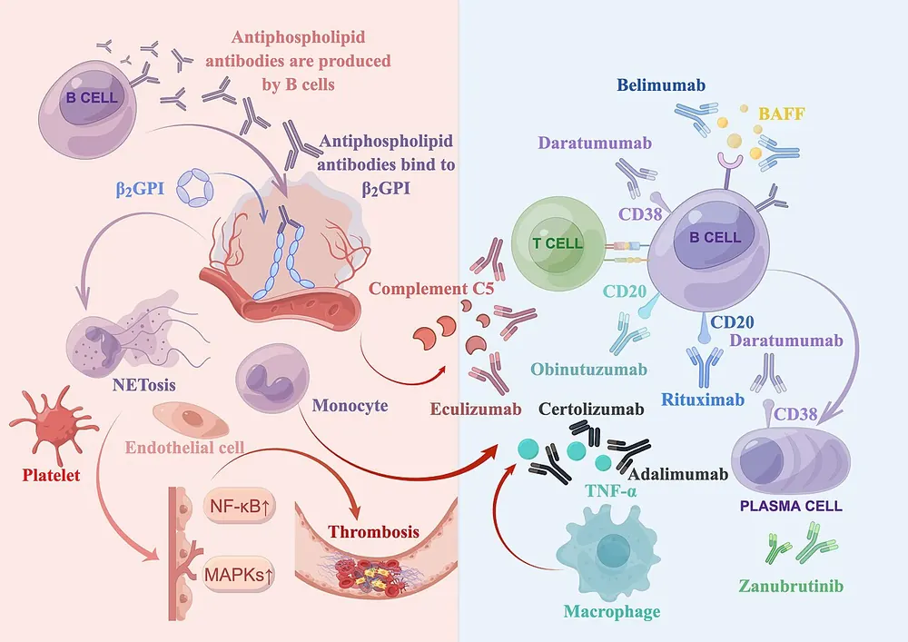
In APS, the immune system produces antiphospholipid antibodies, which actually target proteins that bind to phospholipids, especially beta-2 glycoprotein I. The three main antibodies tested are:
- Lupus anticoagulant
- Anticardiolipin antibodies
- Anti-beta-2 glycoprotein I antibodies
These antibodies promote clotting through several mechanisms:
- Activation of endothelial cells, which start expressing tissue factor and adhesion molecules, making vessel walls "sticky"
- Activation of platelets, increasing clumping
- Activation of monocytes to release tissue factor
- Interference with natural anticoagulants, including protein C and annexin A5, a protein that normally shields phospholipids from clotting factors
- Activation of complement, part of the immune system, which amplifies inflammation and clotting
- Neutrophil extracellular traps (NETs), which provide a scaffold for clots
A "second hit", such as surgery, pregnancy or infection, often triggers a clot in someone with antibodies.
Pregnancy complications
In the placenta, antibodies damage the cells (trophoblasts) that invade the womb lining, impair formation of placental blood vessels, activate complement and promote clots in placental vessels. This reduces blood flow to the baby, leading to miscarriage, stillbirth, growth restriction and pre-eclampsia.
Why the symptoms happen
- Swollen, painful leg results from deep vein thrombosis.
- Breathlessness and chest pain occur when a clot travels to the lungs.
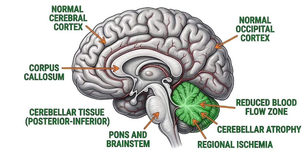
- Stroke or transient ischaemic attack symptoms result from arterial clots in the brain.
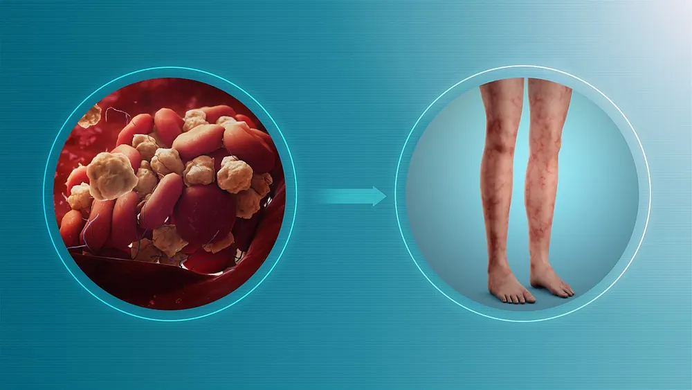
- Lace-like purple rash (livedo reticularis) reflects sluggish blood flow in small skin vessels.
- Low platelets can result from platelet activation and consumption.
How treatment acts on the mechanism
Warfarin reduces the production of vitamin K-dependent clotting factors, preventing new clots. Heparin enhances antithrombin activity and is safe in pregnancy. Low-dose aspirin reduces platelet activation, particularly in pregnancy. Direct oral anticoagulants are generally avoided in high-risk APS because they are less effective at preventing arterial clots. Hydroxychloroquine may reduce antibody effects, especially in lupus.
Types
- Primary APS: no other autoimmune disease
- Secondary APS: with lupus or another autoimmune condition
- Thrombotic APS: clots in veins or arteries
- Obstetric APS: pregnancy complications
- Catastrophic APS: rare, life-threatening multiple organ clots over days
Signs and symptoms
- Pain, swelling and redness in a leg (DVT)
- Sudden breathlessness or chest pain (pulmonary embolism)
- Stroke or mini-stroke symptoms: weakness, numbness, speech difficulty
- Heart attack symptoms
- Recurrent miscarriages, stillbirth or premature birth due to pre-eclampsia
- Lacy, mottled skin rash
- Headaches or migraines
- Easy bruising if platelets are low
How it is diagnosed
- Blood tests for lupus anticoagulant, anticardiolipin antibodies and anti-beta-2 glycoprotein I antibodies
- Repeat testing at least 12 weeks later to confirm persistent antibodies
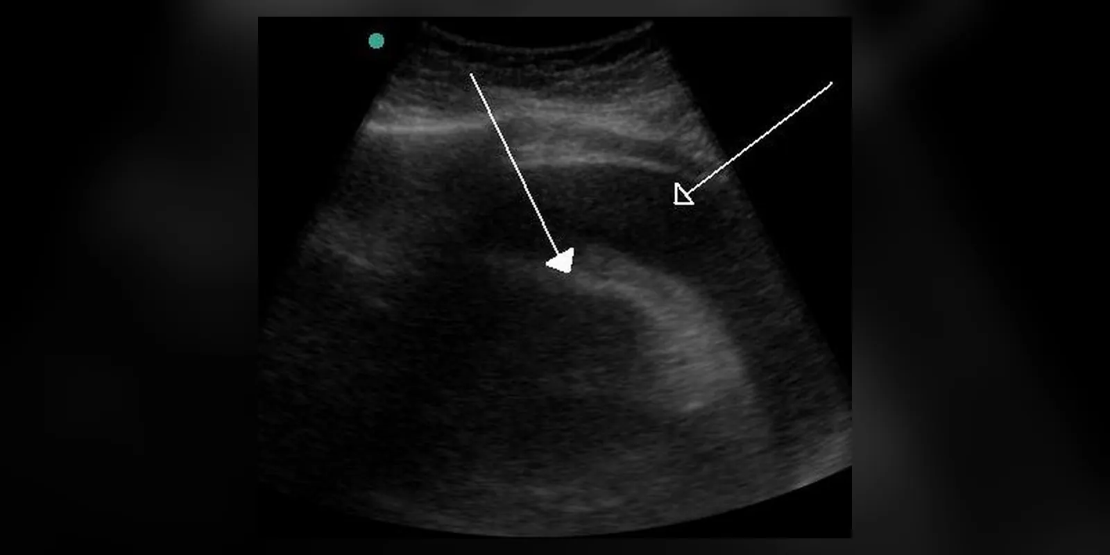
- Imaging for clots: ultrasound, CT pulmonary angiography, brain MRI
- Tests for lupus and other autoimmune diseases
- Full blood count for platelet levels
Possible complications
- Recurrent thrombosis
- Stroke and cognitive problems
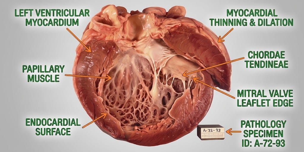
- Heart valve disease
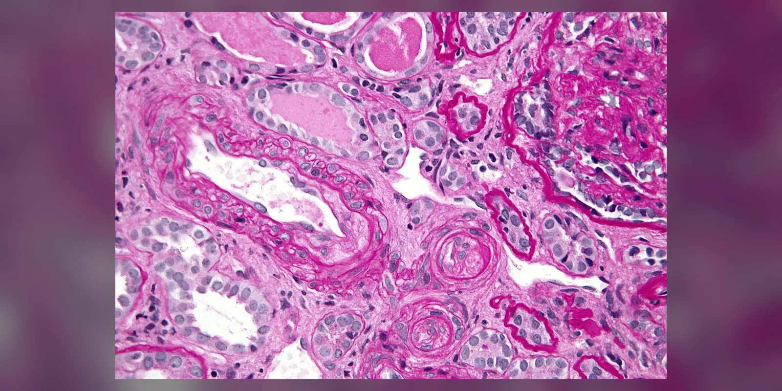
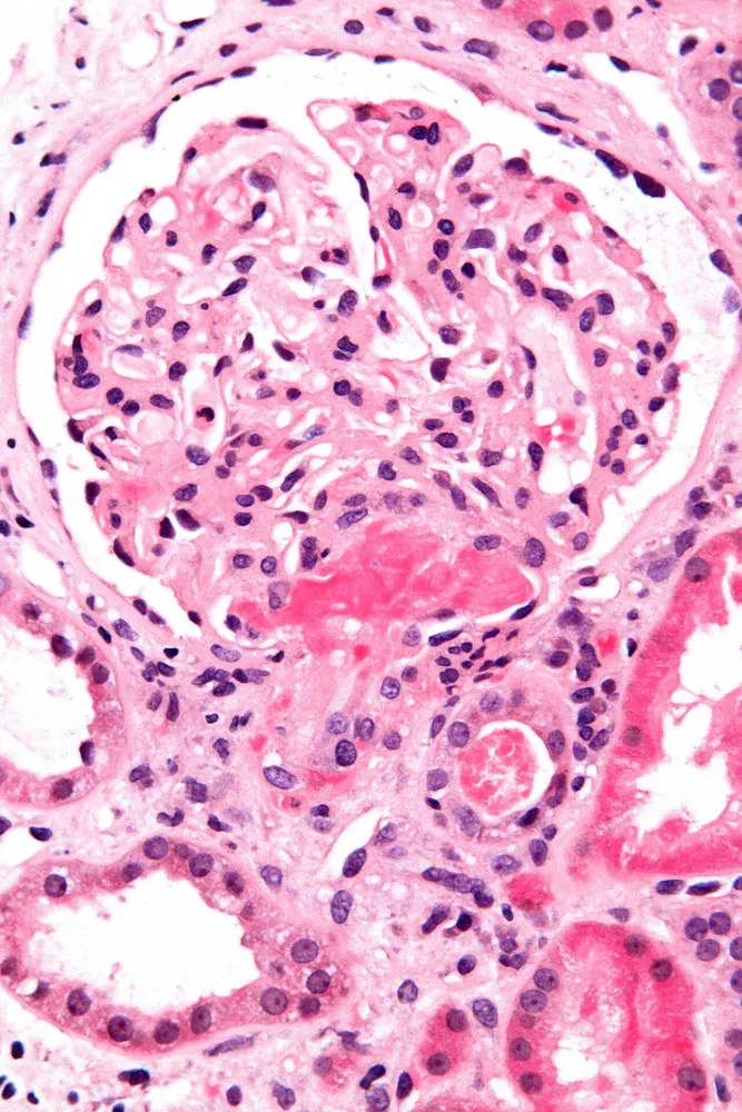
- Kidney damage
- Pulmonary hypertension
- Pregnancy loss and complications
- Catastrophic APS
- Bleeding from anticoagulant treatment
Goals of treatment
Treatment aims to prevent new clots, treat existing clots, and support healthy pregnancies.
Medicines
- Warfarin, long term, for people who have had clots, with a target INR set by the specialist
- Heparin (low molecular weight heparin) in pregnancy and short-term situations
- Low-dose aspirin for some people with antibodies but no clots, and in pregnancy
- Hydroxychloroquine in some cases, especially with lupus
- Catastrophic APS: anticoagulation, steroids, plasma exchange or immunoglobulin
Other treatments
- Specialist pregnancy care with close monitoring
- Compression stockings after DVT
- Avoiding oestrogen-containing contraception and hormone replacement
Living with it and prevention
- Stop smoking
- Stay active and avoid long periods of immobility
- Keep a healthy weight
- Control blood pressure and cholesterol
- Avoid oestrogen-containing contraception
- Discuss clot prevention before surgery, long flights and pregnancy
- Take anticoagulants exactly as prescribed
Monitoring and follow-up
People on warfarin need regular INR checks. Pregnant women with APS have frequent scans to monitor the baby's growth and placental blood flow.
Outlook
With appropriate anticoagulation, most people with APS lead normal lives, and most women with obstetric APS have successful pregnancies with treatment.
Special considerations
- Pregnancy: needs specialist care; warfarin is switched to heparin before six weeks
- People with lupus: higher risk of APS and complications
- Children: APS is rare but can occur