
Overview
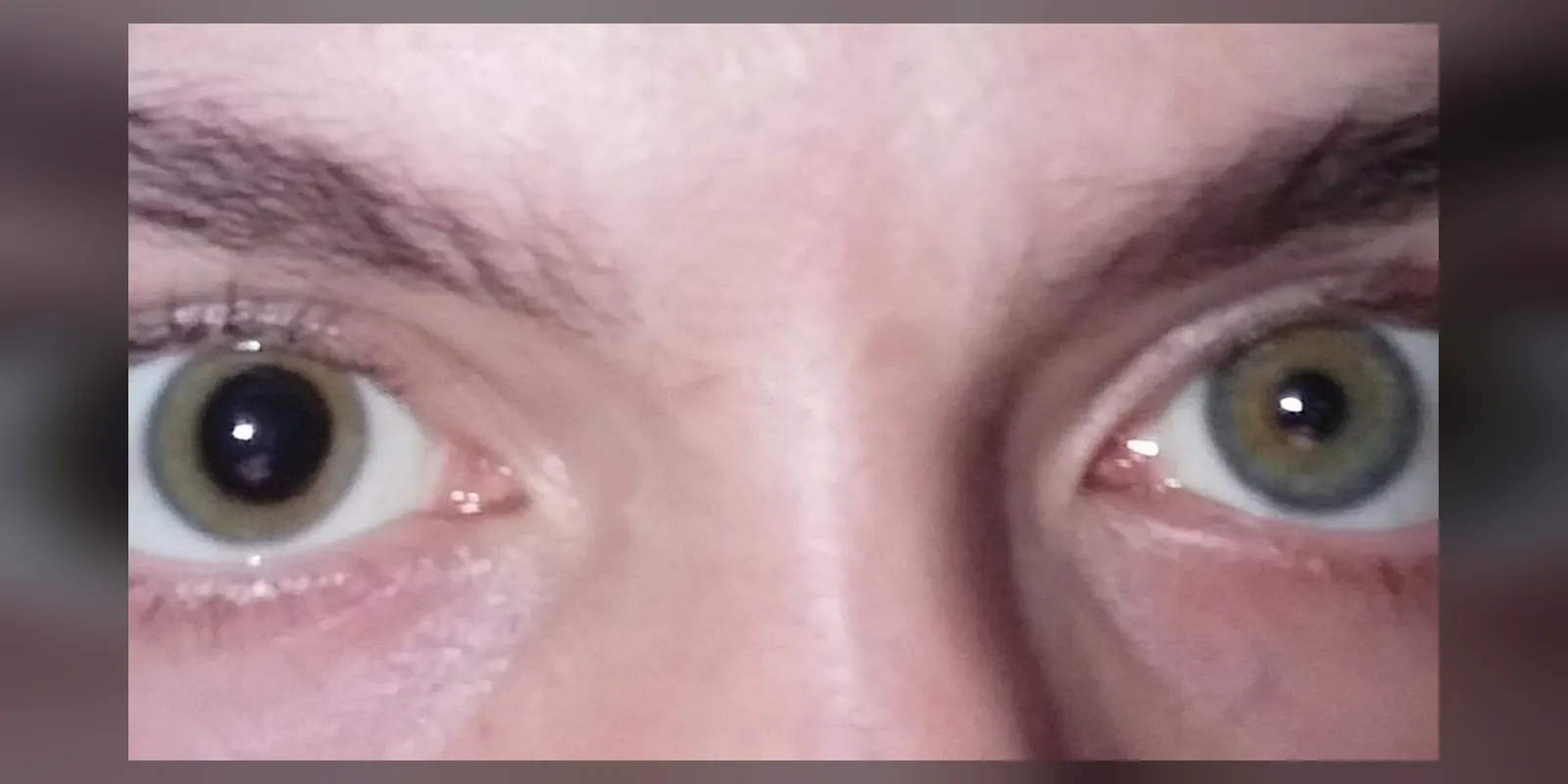
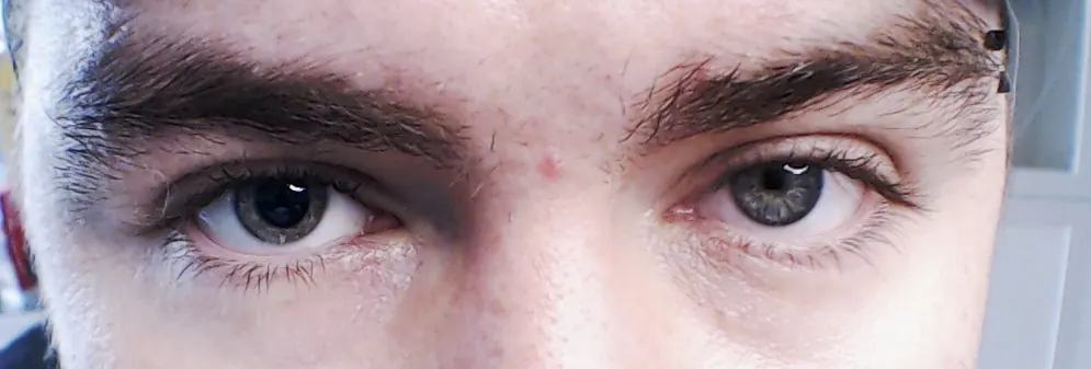
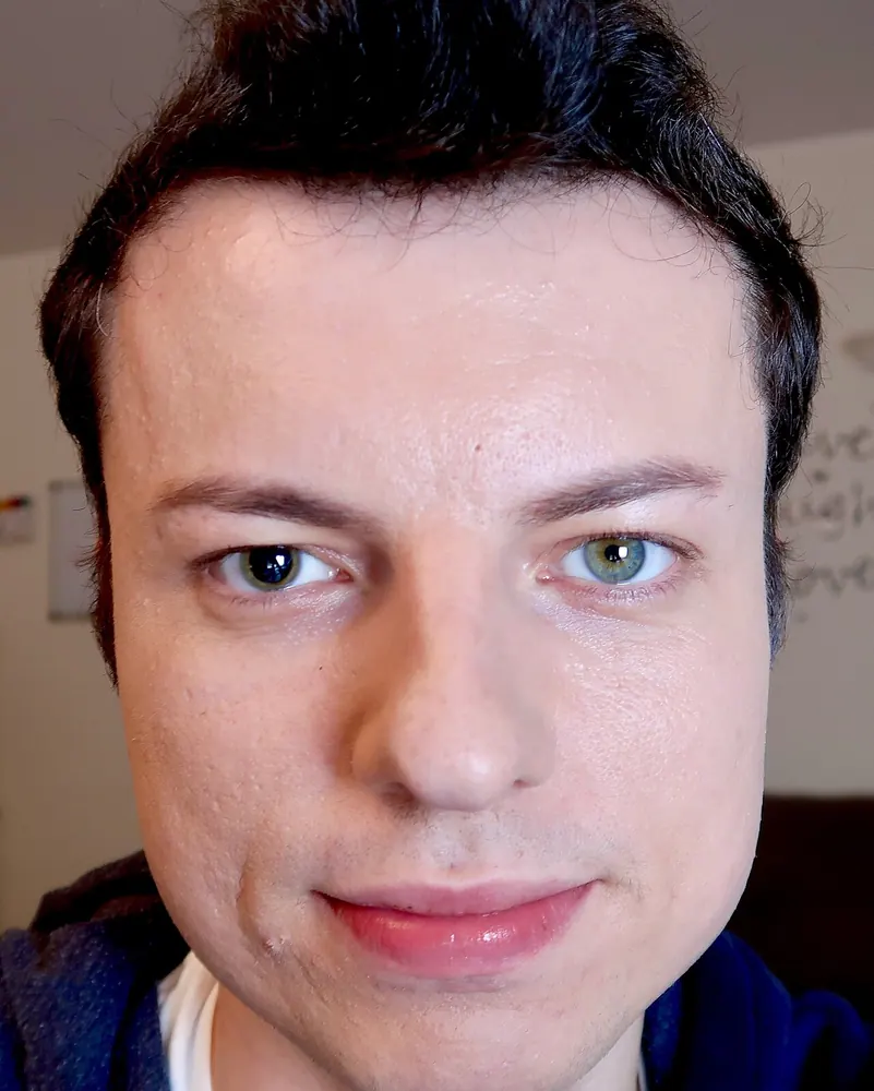
Anisocoria means that the pupils, the black centres of the eyes, are different sizes. A small difference, usually less than 1 millimetre, is common and harmless in about one in five people; this is called physiological anisocoria.
However, new or significant anisocoria can be a sign of a problem with the nerves or muscles that control the pupil, with the eye itself, or with the brain. Causes range from eye drops and injuries to Horner's syndrome, third nerve palsy and Adie's pupil. Sudden anisocoria with symptoms such as drooping eyelid, double vision, headache or confusion needs urgent medical assessment.
When to get urgent help
Seek emergency care for new unequal pupils with a drooping eyelid, double vision, severe headache, neck pain, confusion, weakness, or after a head injury. Also seek urgent care for a painful red eye with blurred vision.
Causes and risk factors
- Physiological anisocoria: harmless, often present for years
- Eye drops or medicines: dilating drops, some inhalers or patches (such as scopolamine), and plant contact (for example angel's trumpet)
- Horner's syndrome: damage to the sympathetic nerve pathway, from carotid artery dissection, lung tumours, stroke or neck surgery
- Third (oculomotor) nerve palsy: from aneurysm, raised pressure in the brain, diabetes or injury
- Adie's tonic pupil: damage to the ciliary ganglion, often after viral illness
- Eye injury or surgery affecting the iris muscles
- Inflammation inside the eye (uveitis)
- Acute angle-closure glaucoma
How it develops (pathophysiology)
Normal pupil control
The pupil size is controlled by two muscles in the iris:
- The sphincter pupillae, a ring of muscle that makes the pupil smaller (constriction). It is controlled by the parasympathetic nervous system via the oculomotor (third cranial) nerve and the ciliary ganglion.
- The dilator pupillae, radial muscle that makes the pupil larger (dilation). It is controlled by the sympathetic nervous system, via a three-neuron pathway that runs from the brain down the spinal cord to the upper chest, then back up along the carotid artery to the eye.
In bright light, the light reflex constricts both pupils equally, because signals from each eye go to both sides of the brainstem. In dim light, both pupils dilate.
What goes wrong
- Physiological anisocoria: a small, stable imbalance in central nerve signals; the difference stays the same in light and dark.
- Horner's syndrome: damage anywhere along the sympathetic pathway weakens the dilator muscle. The affected pupil is smaller, the difference is greater in the dark, and the upper eyelid droops slightly because a small eyelid muscle (Müller's muscle) also loses sympathetic supply. Sweating may be reduced on that side of the face.
- Third nerve palsy: damage to the parasympathetic fibres on the outside of the nerve, such as compression by an aneurysm, weakens the sphincter. The affected pupil is larger and reacts poorly to light, the difference is greater in bright light, and there is usually a drooping eyelid and eye movement problems.
- Adie's tonic pupil: damage to the ciliary ganglion leaves a larger pupil that reacts slowly and poorly to light but constricts slowly when focusing on near objects.
- Pharmacological: drops that block the sphincter (such as atropine) or stimulate the dilator produce a large, unreactive pupil.
Why the symptoms happen
- Unequal pupils result directly from imbalance between the two muscles.
- Light sensitivity or blurred near vision occur with a large pupil.
- Drooping eyelid and double vision occur with third nerve palsy.
- Headache or neck pain may accompany Horner's syndrome from carotid dissection, or third nerve palsy from an aneurysm.
How treatment acts on the mechanism
Treatment depends on the cause: stopping a causative medicine, repairing an aneurysm, treating carotid dissection, managing diabetes, or treating eye disease. Reading glasses or tinted lenses can help symptoms of Adie's pupil.
Types
- Physiological anisocoria
- Anisocoria greater in light (the larger pupil is abnormal): third nerve palsy, Adie's pupil, pharmacological, iris damage
- Anisocoria greater in darkness (the smaller pupil is abnormal): Horner's syndrome, physiological
Signs and symptoms
- Pupils of different sizes, often noticed in photographs or by others
- Light sensitivity
- Blurred vision, especially for near objects
- Drooping eyelid
- Double vision
- Eye pain or redness
- Headache, neck pain or facial pain
- Reduced sweating on one side of the face
How it is diagnosed
- Examination of the pupils in bright and dim light
- Eye movement and eyelid checks
- Slit lamp examination for eye causes
- Eye drop tests: apraclonidine or cocaine drops for Horner's syndrome; dilute pilocarpine for Adie's pupil
- Urgent brain and blood vessel imaging (CT or MR angiography) for suspected third nerve palsy or carotid dissection
- Chest imaging for some causes of Horner's syndrome
Possible complications
Complications depend on the cause, for example stroke from carotid dissection, rupture of an aneurysm, or vision loss from glaucoma.
Goals of treatment
The aim is to identify serious causes quickly and treat the underlying condition.
Medicines
- Treatment of the underlying cause, such as glaucoma treatment, antiplatelet or anticoagulant medicines for carotid dissection, or management of diabetes
- Dilute pilocarpine drops for some people with Adie's pupil
Other treatments
- Neurosurgical or endovascular treatment of aneurysms
- Stopping causative medicines or exposures
- Tinted glasses for light sensitivity
Living with it and prevention
- Wash hands after using medicinal patches or handling plants that affect the pupils
- Wear eye protection to prevent injuries
- Keep old photographs, which help show whether anisocoria is long-standing
Monitoring and follow-up
Physiological anisocoria needs no follow-up. Other causes are monitored according to the underlying condition.
Outlook
Physiological anisocoria is harmless. The outlook for other causes depends on the underlying condition.
Special considerations
- Children: congenital Horner's syndrome can cause a lighter iris colour on the affected side; new Horner's syndrome in a child needs investigation for neuroblastoma
- Head injury: a new dilated pupil may indicate rising pressure in the brain
- Older adults: new anisocoria needs assessment for stroke or aneurysm