
Overview
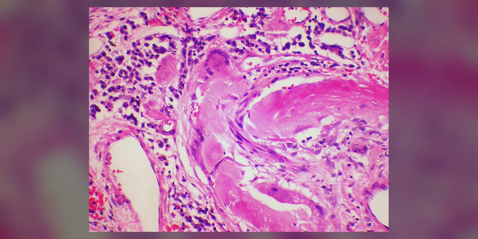
Amyloidosis is a group of rare conditions in which abnormally folded proteins, called amyloid, build up in tissues and organs. Over time, these deposits stiffen and damage organs such as the heart, kidneys, nerves, liver and gut.
There are several types, depending on which protein forms the amyloid. The most common are AL (light chain) amyloidosis, linked to abnormal plasma cells in the bone marrow, and ATTR (transthyretin) amyloidosis, which can be inherited or related to ageing. Symptoms are often vague at first, such as tiredness, swelling, breathlessness or numbness, so diagnosis is often delayed. Treatments now exist that slow or stop amyloid production for many types, making early diagnosis increasingly important.
When to get urgent help
Seek urgent care for severe breathlessness, fainting, chest pain, very low blood pressure, or rapidly increasing swelling.
Causes and risk factors
- AL amyloidosis: abnormal plasma cells produce excess immunoglobulin light chains; sometimes associated with myeloma
- ATTR amyloidosis:
- Wild-type (age-related): mainly affects the heart in older men
- Hereditary (variant): caused by changes in the TTR gene, such as the V122I variant common in people of West African descent
- AA amyloidosis: caused by long-term inflammation, such as rheumatoid arthritis, inflammatory bowel disease, chronic infections or familial Mediterranean fever
- Dialysis-related amyloidosis: beta-2 microglobulin build-up in long-term dialysis
- Localised amyloidosis: confined to one site, such as the skin, bladder or airway
How it develops (pathophysiology)
Normal protein folding
Proteins are long chains of amino acids that fold into precise three-dimensional shapes to work properly. Cells have quality-control systems that refold or destroy misfolded proteins. Some proteins, such as transthyretin (TTR), a carrier protein made by the liver, normally exist as stable groups of four units (tetramers).
What goes wrong
In amyloidosis, a protein misfolds into an abnormal shape rich in beta-pleated sheets. These misfolded molecules stick together:
- Misfolding: the precursor protein becomes unstable. In ATTR, gene variants or ageing make the TTR tetramer fall apart into single units, which misfold. In AL, abnormal light chains made by a plasma cell clone are inherently unstable. In AA, chronic inflammation keeps levels of serum amyloid A protein very high.
- Aggregation: misfolded units clump into small oligomers and then into long, rigid, insoluble amyloid fibrils.
- Deposition: fibrils are deposited outside cells, along with other components such as serum amyloid P and glycosaminoglycans, which stabilise them and resist breakdown.
- Damage: deposits physically distort tissue, stiffen organs and interfere with function. Small oligomers and light chains are also directly toxic to cells.
Organ effects
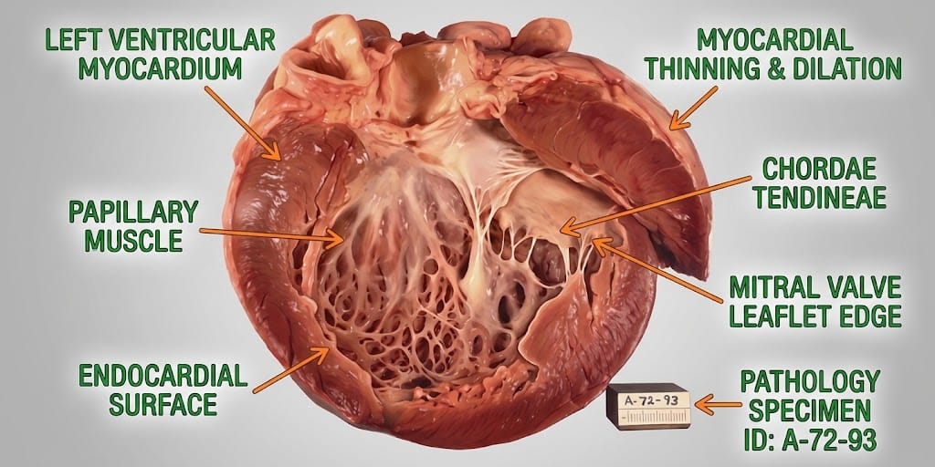
- Heart: amyloid infiltrates heart muscle, making it thick and stiff (restrictive cardiomyopathy). The heart cannot relax and fill properly, causing heart failure, and deposits in the conduction system cause rhythm problems.
- Kidneys: deposits in the glomeruli damage the filter, causing heavy protein loss in urine (nephrotic syndrome) and kidney failure.
- Nerves: deposits damage peripheral and autonomic nerves, causing numbness, pain and blood pressure drops on standing.
- Gut and liver: deposits cause diarrhoea, weight loss and liver enlargement.
- Soft tissues: enlarged tongue, easy bruising (especially around the eyes in AL), carpal tunnel syndrome and spinal stenosis.
How treatment acts on the mechanism
Treatments reduce the supply of the precursor protein:
- AL: chemotherapy and targeted therapies (such as daratumumab, bortezomib) destroy the abnormal plasma cell clone that produces light chains; stem cell transplantation in selected patients.
- ATTR: tafamidis and acoramidis stabilise the TTR tetramer so it does not break apart; gene silencers (patisiran, vutrisiran, inotersen, eplontersen) reduce TTR production by the liver.
- AA: controlling the underlying inflammation lowers serum amyloid A.
Experimental antibodies aim to remove existing deposits.
Types
- AL (light chain) amyloidosis
- ATTR amyloidosis: wild-type or hereditary
- AA amyloidosis
- Dialysis-related amyloidosis
- Localised amyloidosis
- Other rare hereditary types
Signs and symptoms
Symptoms depend on which organs are involved:
- Tiredness and weakness
- Breathlessness and swollen ankles
- Palpitations or fainting
- Foamy urine and swelling
- Numbness, tingling or pain in the hands and feet
- Dizziness on standing
- Diarrhoea or constipation, weight loss
- Enlarged tongue
- Easy bruising, especially around the eyes
- Carpal tunnel syndrome in both hands

How it is diagnosed
- Tissue biopsy stained with Congo red, which shows apple-green birefringence under polarised light; often from abdominal fat, bone marrow or the affected organ
- Typing of amyloid with mass spectrometry or immunohistochemistry
- Blood and urine tests for free light chains and monoclonal proteins
- Heart tests: ECG, echocardiogram, cardiac MRI, troponin and NT-proBNP
- Technetium bone scan (scintigraphy) to diagnose cardiac ATTR without biopsy when blood tests show no light chain abnormality
- Genetic testing for TTR variants
- Kidney and liver tests
Possible complications
- Heart failure and arrhythmias
- Kidney failure
- Nerve damage and disability
- Malnutrition
- Sudden death
- Bleeding
Goals of treatment
The goals are to stop or slow amyloid production, support affected organs, relieve symptoms and improve survival.
Medicines
- AL: chemotherapy combinations, daratumumab, and autologous stem cell transplant for eligible patients
- ATTR: tafamidis or acoramidis (stabilisers); patisiran, vutrisiran, inotersen or eplontersen (gene silencers) for hereditary disease with nerve involvement, and some for cardiomyopathy
- AA: treatment of the underlying inflammatory condition, such as anti-TNF medicines
- Diuretics for fluid overload
- Medicines for nerve pain
Other treatments
- Pacemaker or defibrillator for rhythm problems
- Dialysis or kidney transplantation
- Heart transplantation in selected cases
- Liver transplantation historically for some hereditary ATTR forms
- Nutritional support
- Carpal tunnel surgery
Living with it and prevention
- Seek early assessment for unexplained heart failure, protein in the urine or neuropathy
- Family members of people with hereditary ATTR should consider genetic testing
- Control chronic inflammatory conditions to prevent AA amyloidosis
- Follow fluid and salt advice in heart and kidney involvement
Monitoring and follow-up
Regular monitoring includes light chain levels in AL, heart and kidney function, NT-proBNP, troponin, and imaging. Treatment response is assessed against defined criteria.
Outlook
Outlook depends on the type and the extent of heart involvement. Advanced cardiac AL amyloidosis has a poor prognosis, but early diagnosis and modern treatments have greatly improved survival across types.
Special considerations
- Older men with heart failure, especially with carpal tunnel syndrome, should be considered for wild-type ATTR
- People of West African descent have a higher frequency of the V122I TTR variant
- People on long-term dialysis: dialysis-related amyloidosis